. 2013 Jun;93(6):646-62.
doi: 10.1038/labinvest.2013.55. Epub 2013 Apr 8.

Lack of Cyp1b1 promotes the proliferative and migratory phenotype of perivascular supporting cells

Affiliations

Lack of Cyp1b1 promotes the proliferative and migratory phenotype of perivascular supporting cells

Tammy L Palenski et al. Lab Invest. 2013 Jun.

Abstract

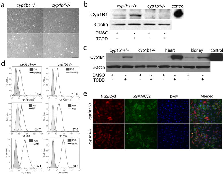
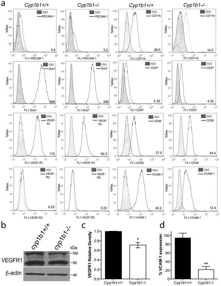
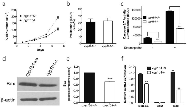
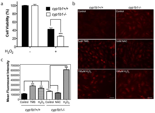
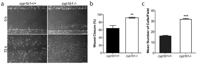
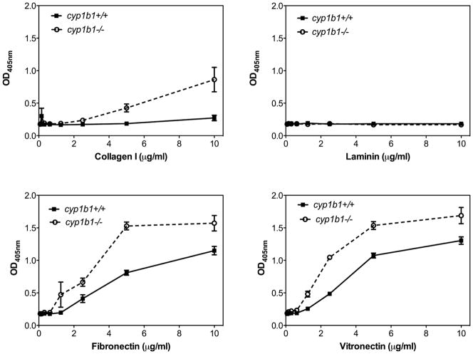
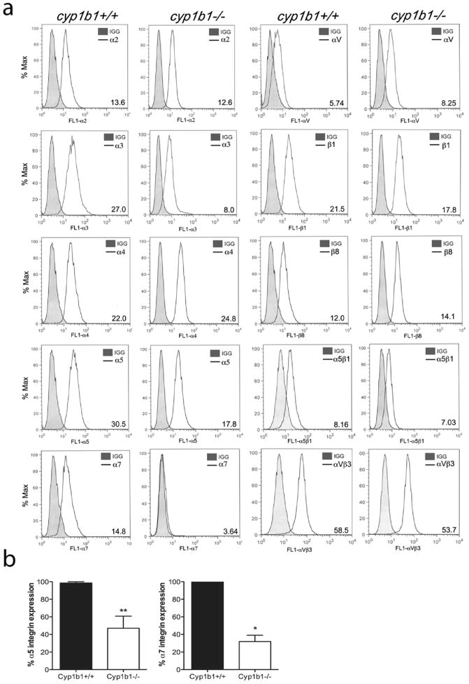
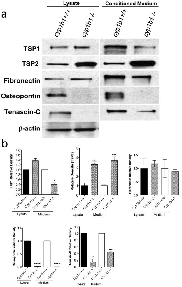
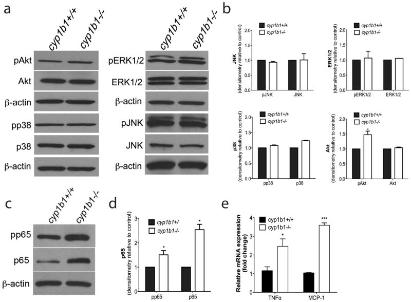
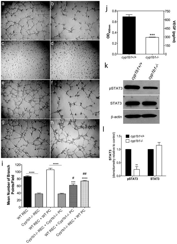
Perivascular supporting cells, including pericytes and smooth muscle cells (PC/SMC), have an integral role during angiogenesis and control vascular remodeling, maturation, and stabilization of neoteric vessels. We recently showed that a Cyp1B1 deficiency in mice results in the attenuation of angiogenesis in vivo and the pro-angiogenic activity of endothelial cells in vitro. However, the contribution of PC/SMC, and more specifically the cell autonomous effects of Cyp1B1 in these processes, needs further investigation. Here we demonstrate that PC constitutively expressed Cyp1B1, and that a deficiency in Cyp1B1 was associated with enhanced proliferation, and decreased apoptosis. Mechanistically, the lack of Cyp1B1 was associated with increased oxidative stress and sustained NF-κB activation, which was reversed by the antioxidant N-acetylcysteine. These changes were also concomitant with alterations in PC migration, adhesion, and expression of various extracellular matrix proteins, including thrombospondin-2. Cyp1B1-deficient PC also expressed decreased levels of vascular endothelial growth factor. Together, our results suggest an important role for Cyp1B1 expression in the regulation of PC proliferation, migration, and survival through modulation of the intracellular oxidative state and NF-κB expression and/or activity. Thus, a lack of Cyp1B1 in PC may have a significant role in vascular dysfunction and integrity, contributing to the attenuation of angiogenesis.


Conflict of interest statement

DISCLOSURE/DUALITYOF INTERST: None

Figures

Figure 1
Figure 1
Isolation and characterization of mouse retinal PC. (a) Cyp1b1+/+and cyp1b1−/− PC were photographed in digital format at ×40 (top panels) and ×100 (bottom panels) magnification. Scale bar in top panel indicates 100 μm; bottom panels 20 μm. (b, c) Cyp1B1 expression in retina, heart, and kidney PC incubated with DMSO or 10nM TCDD for 48 h was evaluated by Western blot analysis of cell lysates (50 μg). Purified recombinant human Cyp1B1 protein was used as a positive control. β-actin was used to assess loading. (d) Cyp1b1+/+and cyp1b1−/− PC were examined for expression of PDGFR-β, NG2, and α-SMA by flow cytometry. Representative mean fluorescent intensities are indicated in bottom right corner of each panel. (e) Indirect immunofluorescent staining using NG2 and αSMA was performed to demonstrate culture purity. DAPI was used to stain cell nuclei. Scale bars indicate 50 μm.
Figure 2
Figure 2
Retinal PC express other vascular cell markers. (a) Expression of PECAM-1, Sca1, VEGF-R1, VEGF-R2, CD11b, CD36, CD47, and VCAM-1 were determined by flow cytometry. Representative mean fluorescent intensities are indicated in bottom right corner of each panel. Shaded areas show staining in the absence of primary antibody. (b) Expression of VEGF-R1 was analyzed by western blot. A representative image is shown. (c) Mean relative density for VEGF-R1 was determined (n=2, *P<0.05). (d) Percent expression of VCAM-1 was determined by flow cytometry (N=3, **P<0.01).
Figure 3
Figure 3
Lack of Cyp1B1 in retinal PC resulted in increased proliferation and decreased apoptosis. (a) The rate of cell proliferation was increased in cyp1b1−/− PC compared to wild-type cells by counting the cell numbers. (b) Cyp1b1+/+ and cyp1b1−/− PC display similar rates of DNA synthesis by flow cytometry (P > 0.05). (c) The rate of apoptosis was determined by measuring caspase activity with luminescent signal from caspase-3/7 DEVD-aminoluciferin substrate. Cyp1b1−/− PC demonstrated a 2 -fold decrease in basal levels of caspase-3/7 and a 2-fold decrease when challenged with 10 nM staurosporine. RLU, relative luminescence unit. (d) Expression of Bax was analyzed by Western blotting. The β-actin level was assessed as a loading control. (e) Quantification of band intensity demonstrated a 1.4-fold decrease in Bax expression in the cyp1b1−/− PC. (**P< 0.001, ***P<0.0001). (f) Relative mRNA expression of Bim-EL, Bax, and Bcl-2 was analyzed by real-time PCR (N=3, **P<0.001)
Figure 4
Figure 4
Cyp1b1−/− retinal PC display higher oxidative stress. (a) Hydrogen peroxide (H2O2) toxicity of retinal PC was measured using the MTS assay. Cyp1b1+/+ and cyp1b1−/− PC were incubated with 150 μM H2O2for 48 h and demonstrated a 1.7 -fold decrease in cell viability (***P < 0.0001). (b) Oxidative stress was measured by dihydroethidium staining in the presence of solvent control DMSO; Cyp1B1 inhibitor, TMS; antioxidant, NAC; and H2O2 for 48 h. Scale bar, 20 μm. (c) Quantitative assessment of mean fluorescent intensity is shown (*P<0.05; ***P<0.0001).
Figure 5
Figure 5
Cyp1b1−/− retinal PC are more migratory. (a) Cell migration was determined by scratch wounding cell monolayers on uncoated tissue culture plates. Wound closure was monitored by photography. Scale bar indicates 100 μm. (b) Quantitative assessment of the data demonstrates an increase in wound closure in the cyp1b1−/− PC (**P<0.001). (c) Transwell migration assays were performed to confirm the migration results. Cyp1b1−/− PC demonstrated a 2 -fold increase in migration (***P < 0.0001).
Figure 6
Figure 6
Cyp1b1−/− retinal PC are more adherent. Adhesion of cyp1b1+/+ and cyp1b1−/− PC to vitronectin, laminin, fibronectin and collagen I was determined as described in Experimental Procedures. Please note an increase in adhesion of cyp1b1/ PC to collagen I (***P<0.0001), fibronectin (**P=0.0021), and vitreonectin (*P=0.021).
Figure 7
Figure 7
Cyp1b1−/− retinal PC exhibit altered expression of integrins. (a) Expression of α1, α2, α3, α4, α5, α7, αV, β1, β8, α5β1, and αVβ3 integrins in cyp1b1+/+and cyp1b1−/− PC was determined by flow cytometry using specific antibodies as described in Materials and methods. The shaded traces show staining in the absence of primary antibody. Representative mean fluorescent intensities are indicated in bottom right corner of each panel. (b) Percent expression of α5 and α7 integrins was determined (N=3, *P<0.05, **P<0.01).
Figure 8
Figure 8
Altered expression of ECM proteins in cyp1b1−/− retinal PC. (a) Cyp1b1+/+ and cyp1b1−/− PC were incubated for 2 days with serum -free PC medium. Cell lysates and conditioned medium were analyzed by Western blot analysis for TSP1, TSP2, fibronectin, osteopontin, and tenascin-C using specific antibodies as described in Materials and methods. (b) Quantitative assessment of band intensity was determined (N=3, *P<0.05, **P<0.001, ***P<0.0001, ****P<0.00001).
Figure 9
Figure 9
Alterations in cellular signaling pathways in cyp1b1−/− retinal PC. (a) Cyp1b1+/+ and cyp1b1−/− PC were analyzed by Western blot analysis for expression of phospho -Akt, total Akt, phospho-p38, total p38, phospho-Erk1/2, total Erk1/2, phospho-JNK, total JNK and β-actin. (b) Quantification of band intensity demonstrated a 1.5 fold increase in phospho-Akt (N=3, *P < 0.05). (c) Levels of phospho-p65 NF-κB, total p65, and β-actin were determined by Western blotting. (d) Quantitative assessment of the data (N=3, *P<0.05). (d, e) Levels of RNA were assessed for NF-κB target genes MCP-1 (***P ≤ 0.0001) and TNFα (*P<0.05).
Figure 10
Figure 10
Loss of Cyp1B1 alters capillary morphogenesis and the production of VEGF through the STAT3 pathway. (a–h) Capillary morphogenesis of retinal EC and PC was assessed by co-culturing cells in Matrigel for 18 hours. Representative images are shown; (a)Wild -type retinal EC, (b) cyp1b1−/− PC, (c) Wild-type PC, (d) cyp1b1−/− PC, (e) Wild-type retinal EC + PC, (f) Cyp1b1−/− retinal EC + cyp1b1−/− PC (g) Wild-type retinal EC + cyp1b1−/− PC (h) Cyp1b1−/− retinal EC + Wild-type PC. Scale bar represents 500 μm.(i) Mean number of branch points were counted (N=3, ***P<0.0001, ****P<0.00001). (j) Analysis of VEGF levels in cyp1b1+/+ and cyp1b1−/− PC demonstrated a 2 -fold decrease (***P ≤ 0.0001). (k) Levels of phospho-STAT3, total STAT3, and β-actin were determined by Western blotting. (l) Quantification of band intensity demonstrated a 4-fold decrease in pSTAT3 (N=3, **P < 0.001).

Similar articles

Cited by

References

    1. Hellstrom M, Kalen M, Lindahl P, Abramsson A, Betsholtz C. Role of PDGF-B and PDGFR-beta in recruitment of vascular smooth muscle cells and pericytes during embryonic blood vessel formation in the mouse. Development. 1999;126:3047–3055. - PubMed
    1. Rolny C, Nilsson I, Magnusson P, et al. Platelet-derived growth factor receptor-beta promotes early endothelial cell differentiation. Blood. 2006;108:1877–1886. - PubMed
    1. von Tell D, Armulik A, Betsholtz C. Pericytes and vascular stability. Exp Cell Res. 2006;312:623–629. - PubMed
    1. Hayden MR, Yang Y, Habibi J, Bagree SV, Sowers JR. Pericytopathy: Oxidative stress and impaired cellular longevity in the pancreas and skeletal muscle in metabolic syndrome and type 2 diabetes. Oxid Med Cell Longev. 2010;3:290–303. - PMC - PubMed
    1. Fleming I. Vascular cytochrome p450 enzymes: physiology and pathophysiology. Trends Cardiovasc Med. 2008;18:20–25. - PubMed

Publication types

Substances