. 2013 Jul;23(7):1182-93.
doi: 10.1101/gr.147314.112. Epub 2013 Apr 8.

Cancer translocations in human cells induced by zinc finger and TALE nucleases

Affiliations

Cancer translocations in human cells induced by zinc finger and TALE nucleases

Marion Piganeau et al. Genome Res. 2013 Jul.

Abstract

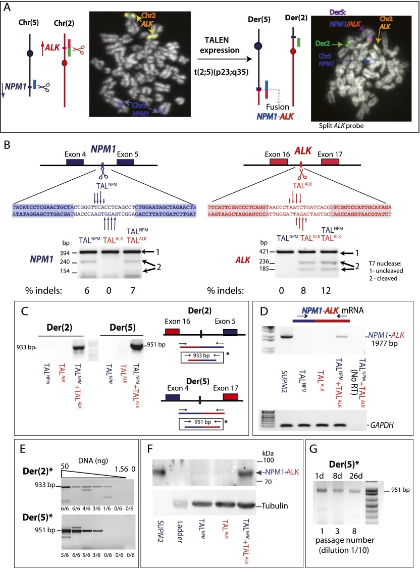
Chromosomal translocations are signatures of numerous cancers and lead to expression of fusion genes that act as oncogenes. The wealth of genomic aberrations found in cancer, however, makes it challenging to assign a specific phenotypic change to a specific aberration. In this study, we set out to use genome editing with zinc finger (ZFN) and transcription activator-like effector (TALEN) nucleases to engineer, de novo, translocation-associated oncogenes at cognate endogenous loci in human cells. Using ZFNs and TALENs designed to cut precisely at relevant translocation breakpoints, we induced cancer-relevant t(11;22)(q24;q12) and t(2;5)(p23;q35) translocations found in Ewing sarcoma and anaplastic large cell lymphoma (ALCL), respectively. We recovered both translocations with high efficiency, resulting in the expression of the EWSR1-FLI1 and NPM1-ALK fusions. Breakpoint junctions recovered after ZFN cleavage in human embryonic stem (ES) cell-derived mesenchymal precursor cells fully recapitulated the genomic characteristics found in tumor cells from Ewing sarcoma patients. This approach with tailored nucleases demonstrates that expression of fusion genes found in cancer cells can be induced from the native promoter, allowing interrogation of both the underlying mechanisms and oncogenic consequences of tumor-related translocations in human cells. With an analogous strategy, the ALCL translocation was reverted in a patient cell line to restore the integrity of the two participating chromosomes, further expanding the repertoire of genomic rearrangements that can be engineered by tailored nucleases.


Figures

Figure 1.
Figure 1.
Induction of t(11;22)(q24;q12) translocations in hES-MP cells with ZFNs. (A) Ewing sarcoma translocations involve breakpoints within the EWSR1 and FLI1 genes on chromosomes 22 and 11, respectively, creating an EWSR1–FLI1 fusion gene on der(22). To induce t(11;22)(q24;q12), ZFNs are expressed in hES-MP cells to create DSBs (scissors) in both genes. (B) ZFNEWS and ZFNFLI cleavage sites are within EWSR1 and FLI1 introns, respectively, relevant to the EWSR1–FLI1 translocation. Zinc fingers in the ZFNs are designed to bind to the shaded sequences. (Arrows) The presumed DSB sites after FokI nuclease domain cleavage. ZFN cleavage activity in hES-MP cells is monitored by a T7-endonuclease assay (Guschin et al. 2010). The region around the ZFN site is amplified; the amplified product is then denatured, reannealed, and then subjected to T7 endonuclease cleavage. Insertions and deletions (indels) characteristic of imprecise DSB repair by NHEJ give rise to T7 endonuclease-cleavable DNA. (C) Nested PCR to detect derivative chromosomes der(11) and der(22) in hES-MP cells. Translocation breakpoint junctions are only detected after expression of both ZFNEWS and ZFNFLI. (D) RT-PCR detection of the EWSR1–FLI1 fusion transcript after ZFNEWS and ZFNFLI expression in hES-MP cells. The forward primer overlaps the exon 2/3 junction of EWSR1, and the reverse primer is within exon 9 of FLI1, amplifying most of the EWSR1–FLI1 coding sequence.
Figure 2.
Figure 2.
Analysis of t(11;22)(q24;q12) breakpoint junctions. (A) Deletion lengths for der(11) and der(22) junctions from ZFN-induced translocations from hES-MP cells. Each value represents the combined deletion from both ends of an individual junction. The median deletion length is indicated by a bar on the graph, and the value is given below the graph. (B) Microhomology length distributions for junctions from ZFN-induced and tumor translocation (Zucman-Rossi et al. 1998). [Der(11) and der(22) are grouped.] Only junctions with simple deletions (i.e., without an insertion) are included. The probability that a junction will have X nucleotides of microhomology by chance assumes an unbiased base composition and is calculated as previously described (Roth et al. 1985). (C) Examples of complex breakpoint junctions with insertions derived from sequences near the DSB sites. Schematics of the derivative and parental chromosomes are shown. The DSB sites on the unrearranged chromosomes are represented by scissors. Segments that are duplicated, deleted, or added are represented as independent blocks and in the orientation relative to that found on the parental chromosomes (see Supplemental Fig. S4 for the sequences).
Figure 3.
Figure 3.
Induction of t(2;5)(p23;q35) translocations with TALENs. (A) ALCL translocations have breakpoints within the NPM1 and ALK genes on chromosomes 2 and 5, respectively, creating an NPM1–ALK fusion gene on der(5). To induce t(2;5)(p23;q35), TALENs are expressed to create DSBs (scissors) in both genes. FISH demonstrates the t(2;5)(p23;q35) translocation after TALEN expression in RPE-1 cells. Red and green signals are from an ALK probe that “breaks apart” upon translocation. The blue signal is from an NPM1 probe. Of 70 metaphases screened, two exhibited translocations and three showed breaks with the ALK break-apart probe, likely due to remaining TALEN expression at this time. (B) TALNPM and TALALK cleavage within NPM1 and ALK introns, respectively, relevant to the NPM1–ALK translocation. DNA binding domains of TALENs are designed to bind the shaded sequences. (Arrows) DSB sites with different tail lengths representing the efficiency of cleavage in vitro, as assayed by in vitro expression of the TALENs (see Supplemental Fig. S6). TALEN cleavage activity in Jurkat cells is monitored by the T7-endonuclease assay, as shown below the sequences. (C) Nested PCR to detect derivative chromosomes der(2) and der(5) in Jurkat cells. Translocation breakpoint junctions are only detected after expression of both TALNPM and TALALK. (D) RT-PCR detection of the NPM1–ALK fusion transcript after TALNPM and TALALK expression in Jurkat cells and in ALCL cell line SUP-M2. The forward primer is within exon 2 of NPM1, and the reverse primer is within exon 29 of ALK, amplifying most of the NPM1-ALK coding sequence. (E) Single-round PCR to detect derivative chromosomes der(2) and der(5) in Jurkat cells. Translocation breakpoint junctions are detected after expression of both TALNPM and TALALK by PCR of the fragment marked (*) in C on serial dilutions of genomic DNA (50, 25, 12.5, 6.25, 3.125, and 1.56 ng). The number of times the PCR was positive for each dilution from six total experiments is indicated. The markers for 933 and 951 bp correspond to der(2) and der(5) junctions, respectively, without end modification. The larger and smaller fragments seen in some of the lanes likely correspond to junctions with large insertions or deletions. (F) Detection of the NPM1–ALK fusion protein in Jurkat cells after TALNPM and TALALK coexpression and in the ALCL cell line SUPM2. The signal from 1 μg of cell extract from SUPM2 cells was compared with 40 μg from Jurkat cells. (G) Expanding pools of RPE-1 cells treated with TALENs. The PCR product corresponding to der(5) was detected when cells were split 1 to 10 every 3 d. The number of passages at each time point is indicated below the gel.
Figure 4.
Figure 4.
Reversion of the t(2;5)(p23;q35) translocation in SUDHL-1 cells. (A) Patient-derived SUDHL-1 cells carry the t(2;5)(p23;q35) translocation and express the NPM1–ALK fusion gene from der(5). To reverse the translocation, TALNPM and TALALK are expressed to create DSBs in both fusion genes; repair between the derivative chromosomes restores intact chromosomes 5 and 2. (B) TALNPM and TALALK efficiently cleave target loci in SUDHL-1 cells. Cleavage is monitored by the T7-endonuclease assay directed to the NPM1 (left) and ALK (right) loci. (C) PCR detection of revertant chromosomes 2 and 5. Reversion is only detected after expression of both TALNPM and TALALK. Because the TALENs cleave to the side of the translocation breakpoint junctions in the SUDHL-1 cells, segments of the other chromosome remain to “tag” the revertant chromosomes to distinguish them from chromosomes that did not participate in the translocation. (D) Sequences of representative revertant chromosomes 5 and 2, which restore the NPM1 and ALK genes, respectively.

References

    1. Barberi T, Willis LM, Socci ND, Studer L 2005. Derivation of multipotent mesenchymal precursors from human embryonic stem cells. PLoS Med 2: e161. - PMC - PubMed
    1. Brunet E, Simsek D, Tomishima M, DeKelver R, Choi VM, Gregory P, Urnov F, Weinstock DM, Jasin M 2009. Chromosomal translocations induced at specified loci in human stem cells. Proc Natl Acad Sci 106: 10620–10625 - PMC - PubMed
    1. Carroll D 2011. Genome engineering with zinc-finger nucleases. Genetics 188: 773–782 - PMC - PubMed
    1. Chansky HA, Barahmand-Pour F, Mei Q, Kahn-Farooqi W, Zielinska-Kwiatkowska A, Blackburn M, Chansky K, Conrad EU III, Bruckner JD, Greenlee TK, et al. 2004. Targeting of EWS/FLI-1 by RNA interference attenuates the tumor phenotype of Ewing’s sarcoma cells in vitro. J Orthop Res 22: 910–917 - PubMed
    1. Colombo E, Martinelli P, Zamponi R, Shing DC, Bonetti P, Luzi L, Volorio S, Bernard L, Pruneri G, Alcalay M, et al. 2006. Delocalization and destabilization of the Arf tumor suppressor by the leukemia-associated NPM mutant. Cancer Res 66: 3044–3050 - PubMed

Publication types