. 2013 May;162(1):333-46.
doi: 10.1104/pp.113.215194. Epub 2013 Apr 8.

Nuclear accumulation of cytosolic glyceraldehyde-3-phosphate dehydrogenase in cadmium-stressed Arabidopsis roots

Affiliations

Nuclear accumulation of cytosolic glyceraldehyde-3-phosphate dehydrogenase in cadmium-stressed Arabidopsis roots

Marco Vescovi et al. Plant Physiol. 2013 May.

Abstract

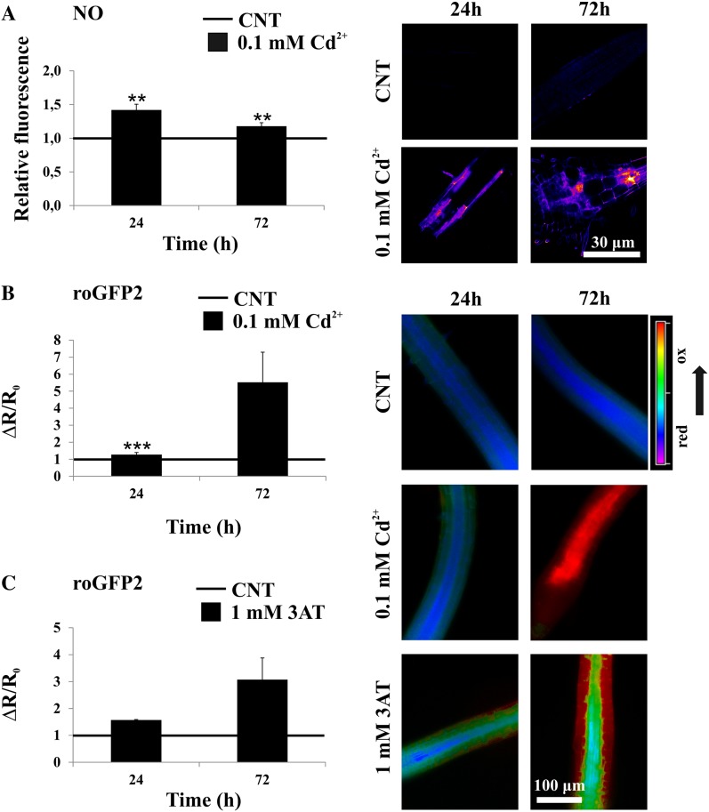
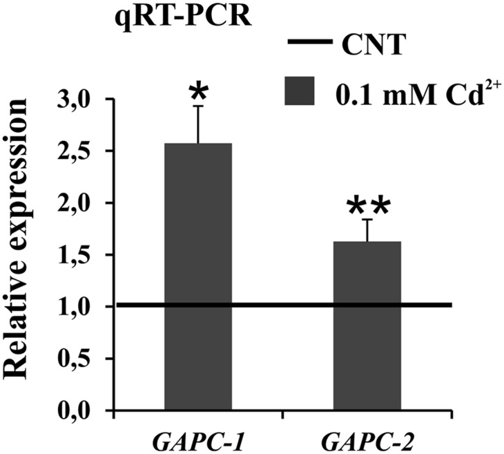
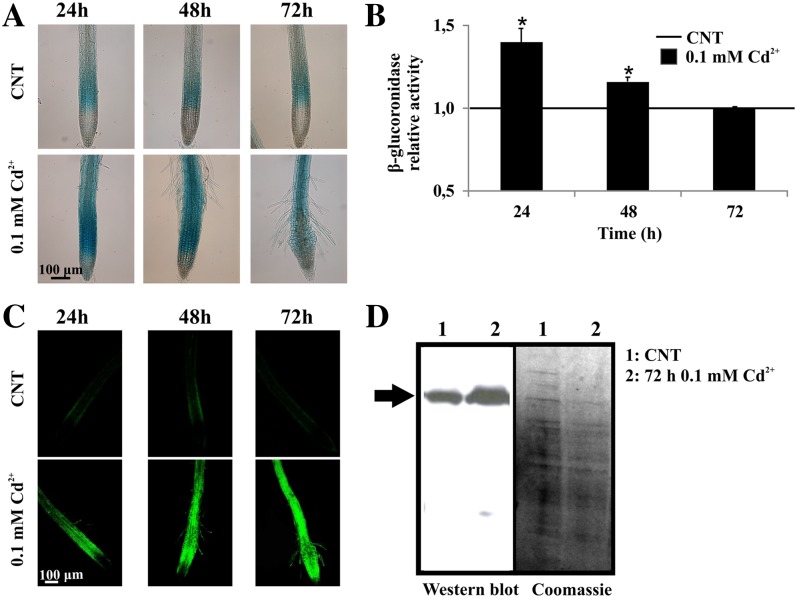
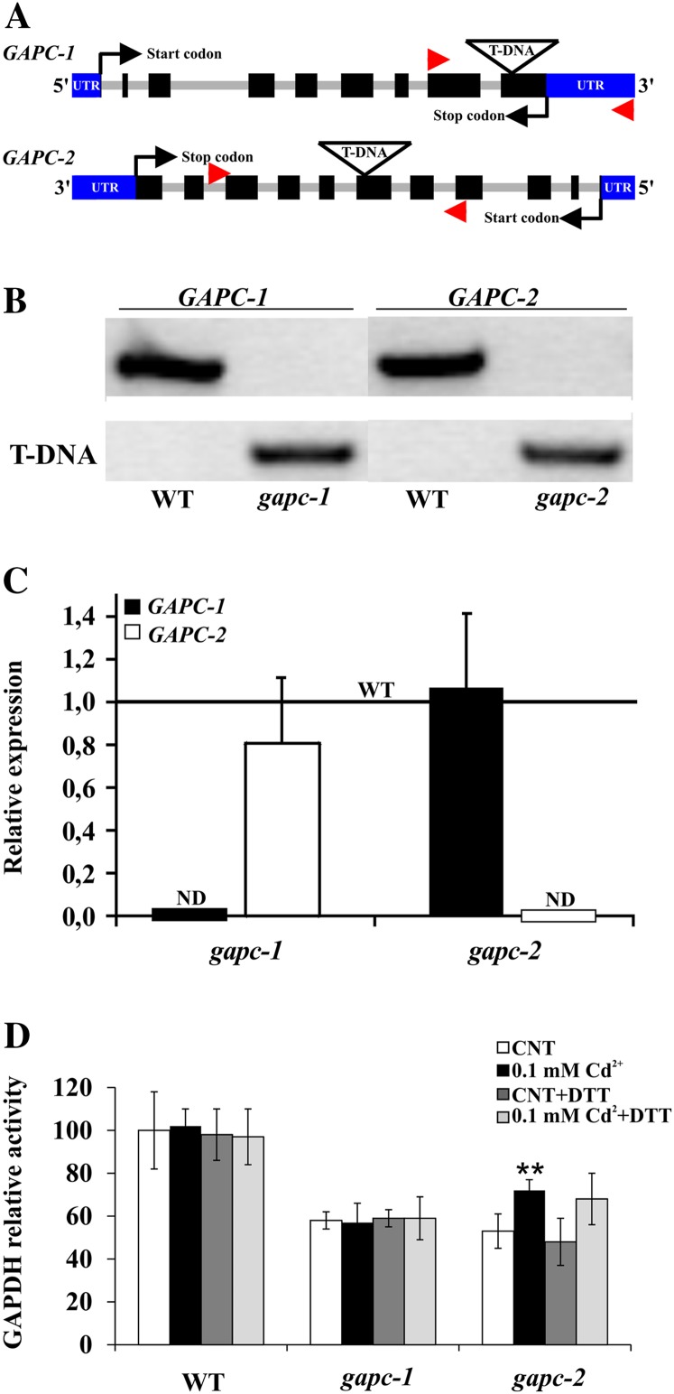
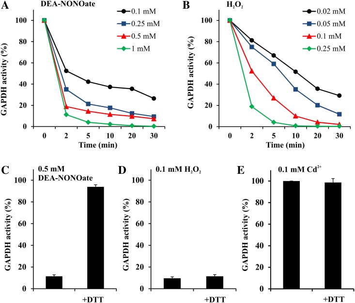
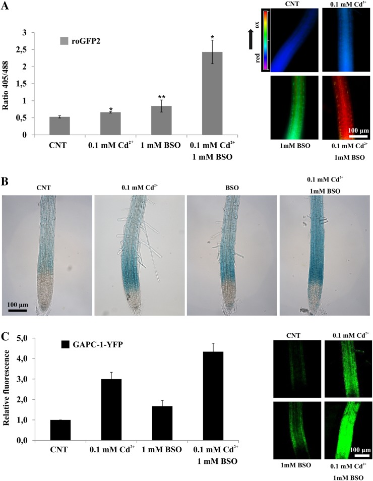
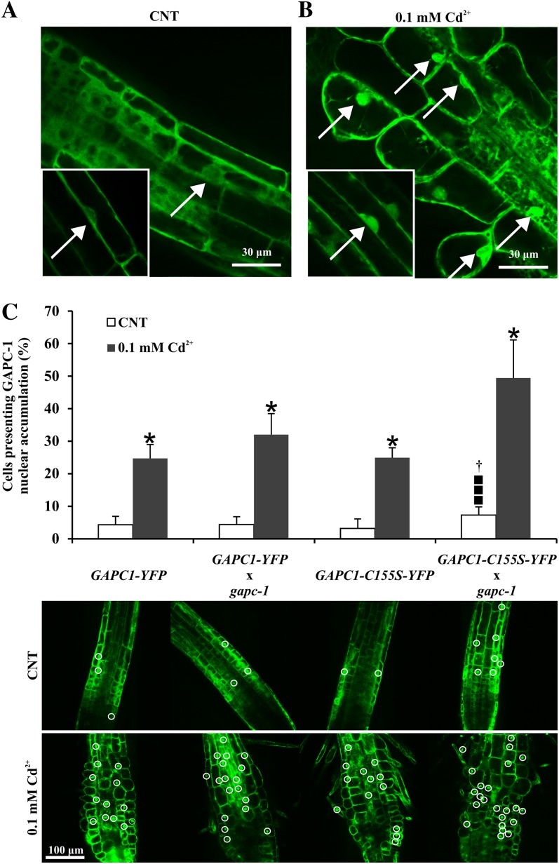
NAD-dependent glyceraldehyde-3-phosphate dehydrogenase (GAPDH) is a ubiquitous enzyme involved in the glycolytic pathway. It has been widely demonstrated that mammalian GAPDH, in addition to its role in glycolysis, fulfills alternative functions mainly linked to its susceptibility to oxidative posttranslational modifications. Here, we investigated the responses of Arabidopsis (Arabidopsis thaliana) cytosolic GAPDH isoenzymes GAPC1 and GAPC2 to cadmium-induced stress in seedlings roots. GAPC1 was more responsive to cadmium than GAPC2 at the transcriptional level. In vivo, cadmium treatments induced different concomitant effects, including (1) nitric oxide accumulation, (2) cytosolic oxidation (e.g. oxidation of the redox-sensitive Green fluorescent protein2 probe), (3) activation of the GAPC1 promoter, (4) GAPC1 protein accumulation in enzymatically inactive form, and (5) strong relocalization of GAPC1 to the nucleus. All these effects were detected in the same zone of the root tip. In vitro, GAPC1 was inactivated by either nitric oxide donors or hydrogen peroxide, but no inhibition was directly provided by cadmium. Interestingly, nuclear relocalization of GAPC1 under cadmium-induced oxidative stress was stimulated, rather than inhibited, by mutating into serine the catalytic cysteine of GAPC1 (C155S), excluding an essential role of GAPC1 nitrosylation in the mechanism of nuclear relocalization, as found in mammalian cells. Although the function of GAPC1 in the nucleus is unknown, our results suggest that glycolytic GAPC1, through its high sensitivity to the cellular redox state, may play a role in oxidative stress signaling or protection in plants.


Figures

Figure 1.
Figure 1.
NO production and redox status in Arabidopsis seedlings exposed to cadmium. A, Seven-day-old Arabidopsis wild-type seedlings treated with 0.1 mm cadmium for 24 and 72 h and stained with DAF-FM diacetate for NO quantification. The relative fluorescence values are related to the fluorescence level in control roots (set to 1). False-colored pictures are representative images of transition and elongation root tip zones considered for the data analyses reported in the graph of A. B, Normalized ratio values (405/488 nm) of cytosolic roGFP2 probe in 7-d-old Arabidopsis roGFP2 transgenic seedlings treated with 0.1 mm cadmium for 24 and 72 h. C, Normalized ratio values (405/488 nm) of cytosolic roGFP2 probe in 7-d-old Arabidopsis roGFP2 transgenic seedlings treated with 1 mm 3-AT for 24 and 72 h. False-colored pictures are representative ratiometric images (405/488-nm ratios) of transition and elongation root tip zones considered for the data analyses reported in the graphs of B and C. Asterisks indicate fluorescence levels or normalized ratios that are significantly different from those found in control roots as calculated by Student’s t test (***P < 0.005 and **P < 0.01).
Figure 2.
Figure 2.
Quantitative real-time RT-PCR expression analysis of GAPC1 and GAPC2 genes in 7-d-old Arabidopsis wild-type seedlings treated with 0.1 mm cadmium for 24 h. The relative expression values of GAPC genes are related to the expression levels in control roots (set to 1). Values represent mean ± sd of relative quantification value of three experiments performed by using templates from three independent biological samples. Asterisks indicate expression levels that are significantly different from those found in control seedlings as calculated by Student’s t test (*P < 0.001 and **P < 0.01).
Figure 3.
Figure 3.
Effects of cadmium treatment on GAPC1 promoter activity and GAPC1-YFP level. A and B, GUS histochemical and quantitative enzymatic activity analyses performed on 7-d-old Arabidopsis pGAPC1::GUS transgenic seedlings treated with 0.1 mm cadmium for 24, 48, and 72 h. The GUS activity assay was performed using total protein extracts from 600 roots in three independent experiments. The relative activity values of GUS are related to the activity in control roots (set to 1). Asterisks indicate GUS activity levels that are significantly different from those found in control roots as calculated by Student’s t test (*P < 0.001). C, Confocal images of 7-d-old Arabidopsis pGAPC1::GAPC1-YFP transgenic seedlings treated with 0.1 mm cadmium for 24, 48, and 72 h. D, Two and one-half micrograms of total protein extracts from roots of 7-d-old Arabidopsis pGAPC1::GAPC1-YFP transgenic seedlings in control conditions or treated with 0.1 mm cadmium for 72 h were assayed by immunoblot analysis using a GFP antibody. Coomassie Blue was used as loading control. The blot was repeated twice.
Figure 4.
Figure 4.
Isolation and characterization of Arabidopsis gapc1 and gapc2 mutants. A, Schemes of GAPC1 and GAPC2 gene structures of gapc1 (SALK_010839) and gapc2 (SALK_016539) insertional mutants. Arrowheads indicate the position of the primers used to screen homozygous mutants. B, PCR analysis of GAPC1 and GAPC2 genes performed on genomic DNA from the wild type and homozygous gapc1 and gapc2 mutants. The upper bands correspond to the amplification of wild-type alleles, and the lower bands correspond to mutated alleles. C, Quantitative real-time RT-PCR expression analysis of GAPC1 and GAPC2 genes in Arabidopsis wild-type and homozygous gapc1 and gapc2 mutant seedlings. The relative expression values are related to the expression levels of the same genes in the wild-type background, which were set to 1. Note that in control conditions the GAPC2 is expressed 100 times more than GAPC1 (Supplemental Fig. S4). Values represent mean ± sd of relative quantification value of three experiments performed by using templates from three independent biological samples. ND, Not detected. D, NADH-specific cytosolic GAPDH catalytic activity analyses performed with total protein extracts from 7-d-old Arabidopsis wild-type, gapc1, and gapc2 mutant seedlings treated with 0.1 mm cadmium for 72 h. Protein extracts were also treated with 10 mm DTT for 15 min and reported in the same graph. The relative activity values of control mutant seedlings and wild-type treated seedlings are related to the activity in control wild-type seedlings, which was set to 100 (corresponding to 418 ± 42 nmol min–1 mg–1). Asterisks indicate activity levels that are significantly different from those found in control seedlings as calculated by Student’s t test (**P < 0.01). [See online article for color version of this figure.]
Figure 5.
Figure 5.
Oxidant treatments of recombinant GAPC1. A, Time- and concentration-dependent inactivation of recombinant GAPC1 by the NO donor DEA-NONOate. B, Time- and concentration-dependent inactivation of recombinant GAPC1 by H2O2. C, Reversibility of DEA-NONOate treatment. Recombinant GAPC1 was incubated for 10 min with 0.5 mm DEA-NONOate, and the reversibility of GAPC1 inactivation was then assessed by incubation for 10 min in the presence of 20 mm DTT. D, Reversibility of H2O2 treatment. Recombinant GAPC1 was incubated for 10 min with 0.1 mm H2O2, and the reversibility of GAPC1 inactivation was then assessed by incubation for 10 min in the presence of 20 mm DTT. E, Effect of cadmium treatment on GAPC1 activity. Recombinant GAPC1 was incubated with 0.1 mm cadmium for 10 min. The reversibility of the treatment was assessed by incubating the protein with 20 mm DTT after exposure to 0.1 mm cadmium for 10 min. Aliquots of the incubation mixtures were withdrawn at the indicated time points, and the NADH-dependent activity was determined. Data are represented as the mean percentage of maximal control activity ± sd. [See online article for color version of this figure.]
Figure 6.
Figure 6.
Effects of inhibition of glutathione biosynthesis on redox status, GAPC1 promoter activity, and GAPC1-YFP level in control and cadmium-treated roots. A, Ratio values (405/488 nm) of 7-d-old Arabidopsis roGFP2 transgenic seedling root tips in control conditions or treated with 0.1 mm cadmium, 1 mm BSO, and 0.1 mm cadmium plus 1 mm BSO for 24 h. False-colored pictures are representative ratiometric images (405/488-nm ratios) of transition and elongation root tip zones considered for the data analyses reported in the graph. B, Histochemical analysis of GUS activity in 7-d-old Arabidopsis pGAPC-1::GUS transgenic seedling root tips in control conditions or treated with 0.1 mm cadmium, 1 mm BSO, and 0.1 mm cadmium plus 1 mm BSO for 24 h. C, YFP fluorescence of 7-d-old Arabidopsis pGAPC-1::GAPC1-YFP transgenic seedling root tips in control conditions or treated with 0.1 mm cadmium, 1 mm BSO, and 0.1 mm cadmium plus 1 mm BSO for 24 h. The graph reports the semiquantitative pixel intensity analysis. The relative fluorescence values are related to the fluorescence level in control roots (set to 1). Pictures are representative images of transition and elongation root tip zones considered for the data analyses reported in the graph. Asterisks indicate fluorescence levels that are significantly different from those found in control roots as calculated by Student’s t test (*P < 0.005 and **P < 0.05).
Figure 7.
Figure 7.
GAPC1-YFP chimeric protein accumulation in the nucleus of cadmium-treated root cells. A, Cells from the elongation zone of a typical root tip of 7-d-old Arabidopsis showing the cytosolic localization of GAPC1-YFP chimeric protein. Inset, representative cell where the YFP signal clearly surrounds the nucleus but is not inside the nucleus. B, Cells from the elongation zone of a typical root tip of 7-d-old Arabidopsis challenged with 0.1 mm cadmium for 72 h showing the characteristic morphological alteration and the GAPC1-YFP accumulation in the nuclei of the responding cells. Inset, representative cell (same focal plane of the inset shown in A) where the YFP signal is inside the nucleus. Note the presence of ectopic root hairs as typical feature of cadmium treatment. C, Effect of mutation of Cys-155 on movement of the protein into the nucleus. Seven-day-old Arabidopsis pGAPC1::GAPC1-YFP, pGAPC1::GAPC1-YFP × gapc1, pGAPC1::GAPC1-C155S-YFP, and pGAPC1::GAPC1-C155S-YFP × gapc1 transgenic seedlings were treated with 0.1 mm cadmium for 72 H, and root tips were analyzed by means of confocal microscope. White circles indicate cells that present GAPC1-YFP nuclear accumulation. In the graph, values represent mean ± sd of the percentages of cells presenting GAPC1-YFP nuclear accumulation. Asterisks indicate values that are significantly different from those found in control roots as calculated by Student’s t test (*P < 0.001). Black squares indicate values that are significantly different from those found in the pGAPC1::GAPC1-YFP × gapc1 transgenic line as calculated by Student’s t test (P < 0.05). Dagger indicates values that are significantly different from those found in pGAPC1::GAPC1-C155S-YFP transgenic line as calculated by Student’s t test (P < 0.001).

Similar articles

Cited by

References

    1. Alonso JM, Stepanova AN, Leisse TJ, Kim CJ, Chen H, Shinn P, Stevenson DK, Zimmerman J, Barajas P, Cheuk R, et al. (2003) Genome-wide insertional mutagenesis of Arabidopsis thaliana. Science 301: 653–657 - PubMed
    1. Anderson LE, Ringenberg MR, Carol AA. (2004) Cytosolic glyceraldehyde-3-P dehydrogenase and the B subunit of the chloroplast enzyme are present in the pea leaf nucleus. Protoplasma 223: 33–43 - PubMed
    1. Apel K, Hirt H. (2004) Reactive oxygen species: metabolism, oxidative stress, and signal transduction. Annu Rev Plant Biol 55: 373–399 - PubMed
    1. Astier J, Rasul S, Koen E, Manzoor H, Besson-Bard A, Lamotte O, Jeandroz S, Durner J, Lindermayr C, Wendehenne D. (2011) S-nitrosylation: an emerging post-translational protein modification in plants. Plant Sci 181: 527–533 - PubMed
    1. Bedhomme M, Adamo M, Marchand CH, Couturier J, Rouhier N, Lemaire SD, Zaffagnini M, Trost P. (2012) Glutathionylation of cytosolic glyceraldehyde-3-phosphate dehydrogenase from the model plant Arabidopsis thaliana is reversed by both glutaredoxins and thioredoxins in vitro. Biochem J 445: 337–347 - PubMed

Publication types

MeSH terms