. 2013 Jul;62(7):2471-80.
doi: 10.2337/db12-1001. Epub 2013 Apr 8.

Conversion of mature human β-cells into glucagon-producing α-cells

Affiliations

Conversion of mature human β-cells into glucagon-producing α-cells

H Siebe Spijker et al. Diabetes. 2013 Jul.

Abstract

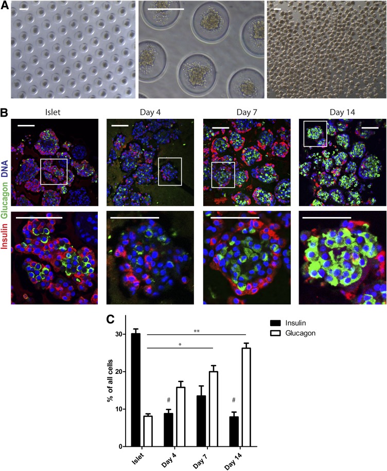
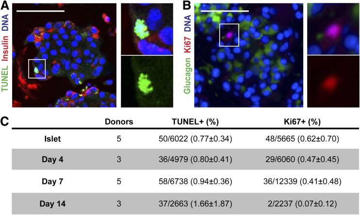
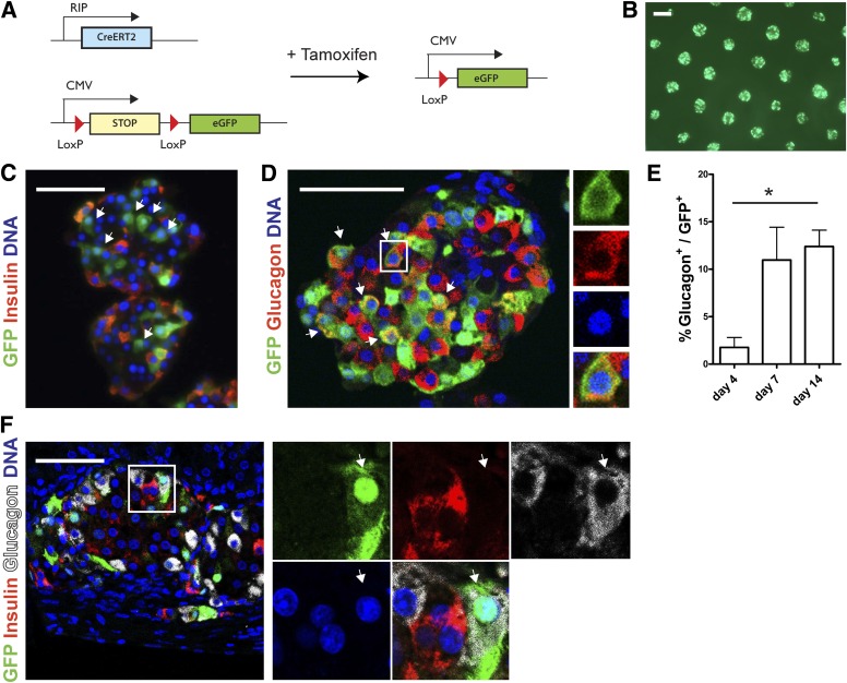
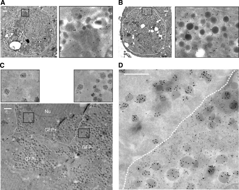
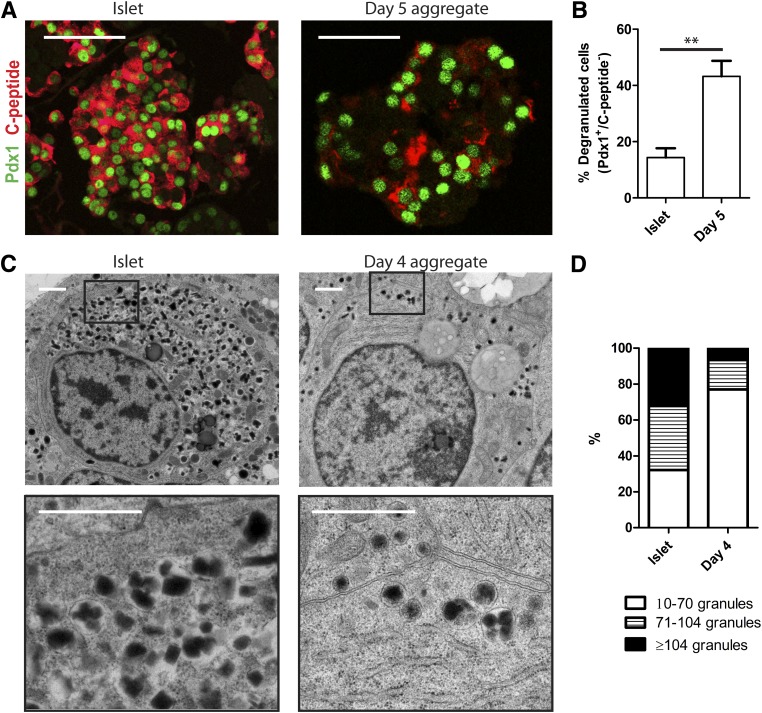
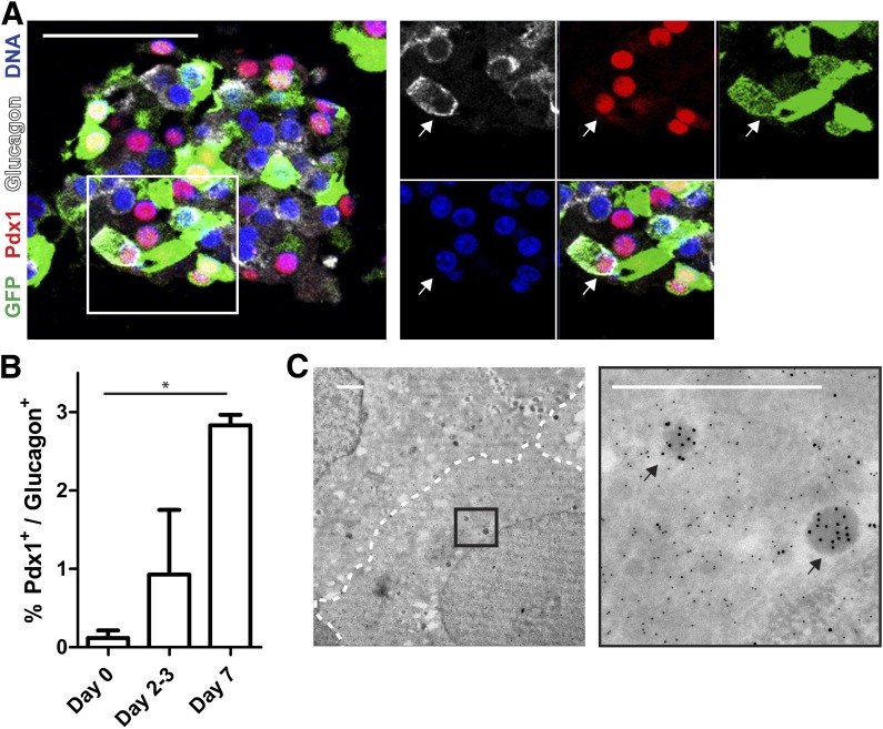
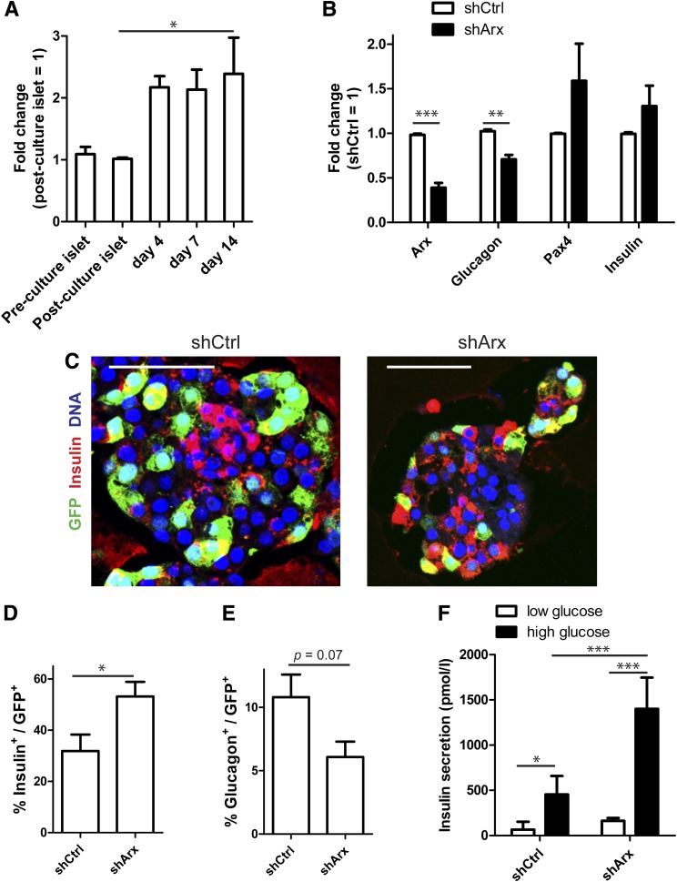
Conversion of one terminally differentiated cell type into another (or transdifferentiation) usually requires the forced expression of key transcription factors. We examined the plasticity of human insulin-producing β-cells in a model of islet cell aggregate formation. Here, we show that primary human β-cells can undergo a conversion into glucagon-producing α-cells without introduction of any genetic modification. The process occurs within days as revealed by lentivirus-mediated β-cell lineage tracing. Converted cells are indistinguishable from native α-cells based on ultrastructural morphology and maintain their α-cell phenotype after transplantation in vivo. Transition of β-cells into α-cells occurs after β-cell degranulation and is characterized by the presence of β-cell-specific transcription factors Pdx1 and Nkx6.1 in glucagon(+) cells. Finally, we show that lentivirus-mediated knockdown of Arx, a determinant of the α-cell lineage, inhibits the conversion. Our findings reveal an unknown plasticity of human adult endocrine cells that can be modulated. This endocrine cell plasticity could have implications for islet development, (patho)physiology, and regeneration.


Figures

FIG. 1.
FIG. 1.
Human islet cell aggregate formation results in an increase in glucagon+ cells. A: Culture of dispersed islet cells in microwells (diameter 200 μm) results in homogenously sized islet cell aggregates that remain intact after flushing them out of the microwells. Scale bars: 200 μm. B: Human islet cell aggregate formation results in a distinct architecture after reaggregation: β-cells (insulin, red) are located at the rim and α-cells in the center (glucagon, green). Scale bars: 100 μm. C: Over time, an increased proportion of glucagon+ cells was present in islet cell aggregates. Data are presented as means ± SEM. n = 4–9 donors. *P < 0.05, **P < 0.005 vs. intact islets glucagon; #P < 0.05 vs. intact islets insulin.
FIG. 2.
FIG. 2.
Change in endocrine hormone distribution is not due to apoptosis or proliferation. A and B: Representative picture of TUNEL assay combined with insulin staining (A) and Ki67 staining with glucagon (B). C: Quantification of apoptotic and proliferating cells defined by the number of positive nuclei in counted cells. Data are presented as means ± SEM. n = 3–6 donors.
FIG. 3.
FIG. 3.
Increased number of glucagon+ cells results from the conversion of β-cells. A: Schematic representation of conditional β-cell–specific lineage tracing using two lentiviral vectors. B: Live cell imaging of transduced human islet cell aggregates shows an equal distribution of epifluorescent GFP throughout the microwell chip. C: After 7 days of reaggregation, GFP+ cells are observed that do not express insulin (arrows). D and E: Glucagon+/GFP+ cells (arrows) are present in human islet cell aggregates (D) with increasing numbers over time (E). Data are presented as means ± SEM. n = 3–6 donors. *P < 0.05. F: Glucagon+/GFP+ cell (arrows) in a graft after transplantation under the kidney capsule of NOD/SCID mice. Scale bars = 50 μm, except for B: 200 μm.
FIG. 4.
FIG. 4.
Converted cells are regular α-cells based on ultrastructural morphology. A and B: Immunogold labeling for GFP (15-nm gold particles) is present as black dots in both cells with granules with a typical crystalline structure containing insulin (A) and cells with more homogenous dark and dense noninsulin granules (B). C and D: Double immunogold labeling for GFP (10-nm gold particles) and glucagon (15-nm gold particles) shows the presence of α-cell granules in both GFP+ (converted) and GFP cells (C). Immunogold labeling for GFP is absent within granules (D). The borders between the cells are marked manually by a broken line (Nu = nucleus). All scale bars = 500 nm.
FIG. 5.
FIG. 5.
Reaggregation is accompanied by β-cell degranulation. A: Immunostaining for Pdx1 (green) and C-peptide (red) in intact islets and 5 days after reaggregation. Scale bars = 100 μm. B: Quantification of the number of Pdx1+/C-peptide cells. Data are presented as means ± SEM. n = 3 donors. **P < 0.005. C: Electron microscopy photographs showing insulin granules in intact islet β-cells and 4 days after reaggregation. Scale bars = 1 μm. D: Distribution of the amount of insulin granules per β-cell in intact islets and 4 days after reaggregation (for representation of the distribution, granule number in intact islets was divided in tertiles; >50 cells were counted per condition).
FIG. 6.
FIG. 6.
β- to α-cell transition is marked by the presence of glucagon+ cells containing β-cell transcription factors. A: Immunostaining for GFP (green), Pdx1 (red), and glucagon (white) (arrows in enlargement show triple-positive cell). Scale bar = 100 μm. B: Quantification of Pdx1+/glucagon+ cells shows an increase during reaggregation (n = 3 donors, *P < 0.05). C: Double immunogold labeling for GFP (10-nm gold particles) and glucagon (15-nm gold particles) after reaggregation (arrows show single glucagon granules). Scale bar = 1 μm.
FIG. 7.
FIG. 7.
Arx knockdown inhibits the conversion of β-cells into α-cells. A: Arx gene expression in human islet cell aggregates compared with control intact islets (n = 3–6 donors). B: shRNA directed against Arx (shArx) results in knockdown of Arx mRNA that is accompanied by changes in glucagon, Pax4, and insulin mRNA levels (n = 4 donors). C: Combining β-cell lineage tracing and shRNA treatment shows a large proportion of GFP+/insulin cells in shCtrl-treated islet cell aggregates but an increase in GFP+/insulin+ cells in shArx-treated islet cell aggregates. D and E: The number of remaining β-cells is increased in shArx-treated aggregates compared with shCtrl (D), while the number of converted cells is decreased as represented by GFP+/glucagon+ cells (E) (n = 4 donors). F: Representative graph of a glucose-stimulated insulin secretion assay that shows increased insulin secretion after treatment with shArx (mean ± SD of quadruplicates). All experiments were performed after 7–14 days’ reaggregation. Scale bars = 50 μm. *P < 0.05, **P < 0.005, ***P < 0.0001.

References

    1. Cabrera O, Berman DM, Kenyon NS, Ricordi C, Berggren PO, Caicedo A. The unique cytoarchitecture of human pancreatic islets has implications for islet cell function. Proc Natl Acad Sci USA 2006;103:2334–2339 - PMC - PubMed
    1. Brissova M, Fowler MJ, Nicholson WE, et al. Assessment of human pancreatic islet architecture and composition by laser scanning confocal microscopy. J Histochem Cytochem 2005;53:1087–1097 - PubMed
    1. Bosco D, Armanet M, Morel P, et al. Unique arrangement of alpha- and beta-cells in human islets of Langerhans. Diabetes 2010;59:1202–1210 - PMC - PubMed
    1. Seymour PA, Sander M. Historical perspective: beginnings of the beta-cell: current perspectives in beta-cell development. Diabetes 2011;60:364–376 - PMC - PubMed
    1. Courtney M, Pfeifer A, Al-Hasani K, et al. In vivo conversion of adult α-cells into β-like cells: a new research avenue in the context of type 1 diabetes. Diabetes Obes Metab 2011;13(Suppl. 1):47–52 - PubMed

Publication types

MeSH terms