. 2013 Apr 23;110(17):E1621-30.
doi: 10.1073/pnas.1218984110. Epub 2013 Apr 8.

Bacillus subtilis biofilm induction by plant polysaccharides

Affiliations

Bacillus subtilis biofilm induction by plant polysaccharides

Pascale B Beauregard et al. Proc Natl Acad Sci U S A. .

Abstract

Bacillus subtilis is a plant-beneficial Gram-positive bacterium widely used as a biofertilizer. However, relatively little is known regarding the molecular processes underlying this bacterium's ability to colonize roots. In contrast, much is known about how this bacterium forms matrix-enclosed multicellular communities (biofilms) in vitro. Here, we show that, when B. subtilis colonizes Arabidopsis thaliana roots it forms biofilms that depend on the same matrix genes required in vitro. B. subtilis biofilm formation was triggered by certain plant polysaccharides. These polysaccharides served as a signal for biofilm formation transduced via the kinases controlling the phosphorylation state of the master regulator Spo0A. In addition, plant polysaccharides are used as a source of sugars for the synthesis of the matrix exopolysaccharide. The bacterium's response to plant polysaccharides was observed across several different strains of the species, some of which are known to have beneficial effects on plants. These observations provide evidence that biofilm genes are crucial for Arabidopsis root colonization by B. subtilis and provide insights into how matrix synthesis may be triggered by this plant.


Conflict of interest statement

The authors declare no conflict of interest.

Figures

Fig. 1.
Fig. 1.
B. subtilis cells colonizing A. thaliana roots express matrix genes. Wild-type (3610) cells harboring PtapA-yfp were coincubated with 6-d-old seedlings of A. thaliana and imaged at various time points postinoculation. Shown are overlays of fluorescence (false-colored green) and transmitted light (gray) images. Pictures are representative of at least ten independent roots. Arrows point toward some of the nonfluorescent cells. (Scale bars: 10 μm.)
Fig. 2.
Fig. 2.
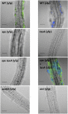
B. subtilis forms biofilms on plant roots in an eps tasA-dependent fashion and requires spo0A and sinI. Wild-type (WT) cells constitutively expressing YFP or CFP and various mutant strains constitutively expressing YFP were coincubated with 6-d-old seedlings of A. thaliana. For the bacterial coinoculation, eps and tasA mutant cells were added at a 1:1 ratio. Colonization of the root was observed after 24 h. Overlays of fluorescence (false colored green for YFP or blue for CFP) and transmitted light images (gray) are shown. Pictures are representative of at least twelve independent roots. (Scale bars: 50 μm.)
Fig. 3.
Fig. 3.
Plant extracts but not root exudates induce biofilm formation. Pellicle formation of wild-type (3610) cells in MSNc with no addition (−) or with root exudates or plant extracts added at the onset of the assay. Images are top-down view of wells and were taken after 24 h at 30 °C. The “No cells” control is shown to highlight the appearance of bacterial cell growth in the inoculated wells.
Fig. 4.
Fig. 4.
Plant polysaccharides promote biofilm formation. (A) Pellicle formation of wild-type cells in MSNc. The indicated di- ,tri-, or polysaccharides were added at a final concentration of 0.5% at the onset of the assay. AG, Arabinogalactan. Images are top-down views of wells and were taken after 24 h at 30 °C. (B) Pellicle weight assay of wild-type cells in MSNc. The indicated di-, tri-, or polysaccharides were added at a final concentration of 0.5% at the onset of the assay. Analysis of variance revealed a significant main group effect between the conditions used [F(10, 75), P < 0.001]. Tukey's post hoc test revealed that arabinogalactan, pectin, and xylan (marked with asterisks) showed greater pellicle mean mass compared with all other conditions. (C) Flow cytometry analysis of wild-type cells harboring the PtapA-yfp reporter grown in MSNc with indicated additions or no supplements (−). Fluorescence intensity in arbitrary units is shown on the x axis and number of cells is shown on the y axis. Results shown are representative of three independent experiments.
Fig. 5.
Fig. 5.
The master regulator Spo0A and the SinI antirepressor are involved in responding to plant polysaccharides. (A) Top-down view of pellicle assay in which the indicated regulatory mutant cells were incubated for 24 h at 30 °C in the presence of arabinogalactan (AG), pectin, or xylan in MSNc medium. “No cells” controls show that mutant cells are able to grow but do not form a pellicle. (B) Weight quantification of pellicles formed in the same conditions as in A. A two-way analysis of variance revealed a significant difference between the mutants and wild type [F(2, 27), P < 0.001]. Tukey's post hoc test revealed that sinI and spo0A (marked with asterisks) showed lower pellicle mean mass compared with wild type in the same conditions.
Fig. 6.
Fig. 6.
KinC and KinD are involved in sensing and responding to plant polysaccharides. (A) Top-down view of pellicle assay in which the indicated regulatory mutant cells were incubated for 24 h at 30 °C in the presence of arabinogalactan (AG), pectin, or xylan in MSNc medium. (B) Weight quantification of pellicles formed in the same conditions as in A. A two-way analysis of variance revealed a significant difference between the mutants marked with an asterisk and wild type [F(6, 63), P < 0.001]. Tukey's post hoc test revealed that kinCD in the presence of AG and pectin (marked with asterisks) showed lower pellicle mean mass compared with wild type in the same conditions, but that was not the case in the presence of xylan.
Fig. 7.
Fig. 7.
Plant polysaccharides provide a substrate that is incorporated into the matrix EPS. (A) A proposed model for the metabolism of galactose that is incorporated into the matrix EPS. GalK and GalT convert exogenous galactose to UDP-galactose, which is used in EPS production. GalE converts UDP-glucose into UDP-galactose, producing UDP-galactose from central metabolism. (B) Top-down view of pellicle assay in which the indicated mutants were incubated for 24 h at 30 °C in the presence of arabinogalactan (AG), pectin, or xylan in MSNc medium. Results are representative of three experiments. (C) Weight quantification of pellicles formed in the same conditions as in B. A two-way analysis of variance revealed a significant difference between the mutants [F(6, 63), P < 0.001]. Tukey's post hoc test revealed that in the presence of AG and pectin, only galE galKT mutant (marked with asterisks) showed lower pellicle mean masses compared with wild type (WT), but in presence of xylan both galE and galE galKT are significantly different from WT. (D) Root colonization of 3610, galE, galKT, and galE galKT mutants constitutively expressing YFP. When indicated, galactose is added at a concentration of 0.05%. Pictures are representative of at least 16 independent roots for those without galactose and 10 for those with galactose. (Scale bars: 50 μm.)
Fig. 8.
Fig. 8.
A. thaliana arabinogalactan proteins induce biofilm formation. (A) Pellicle formation of wild-type cells in MSNc in the presence of 0.05% or 0.5% commercial arabinogalactan (AG) or purified A. thaliana arabinogalactan proteins (A. thaliana AGPs). Images are top-down views of wells and were taken after 24 h at 30 °C. (B) Weight quantification of pellicles formed in the same conditions as in A. A two-way analysis of variance revealed a significant difference between the conditions marked with an asterisk and the untreated sample [F(4, 15), P < 0.001]. Tukey's post hoc test revealed 0.5% AG, and both concentrations of A. thaliana AGPs showed higher pellicle mean mass compared with the untreated well.
Fig. 9.
Fig. 9.
Biofilm formation by plant growth-promoting Bacillus strains is influenced by plant polysaccharides. Pellicle formation by B. subtilis 3610, the plant growth-promoting strain B. subtilis GB03, and B. amyloliquefaciens FZB42 in MSNc with arabinogalactan (AG), pectin, and xylan. Top-down view of pellicle after incubation for 24 h at 30 °C. Results are representative of three experiments.

References

    1. Hiltner L. Über neuere Erfahrungen und Probleme auf dem Gebiete der Bodenbakteriologie unter bessonderer Berücksichtigung der Gründung und Brache [About our experiences and problems in the area of soil bacteriology under special regards of cultivated and uncultivated land] Arbeiten der Deutschen Landwirtschaftlichen Gesellschaft. 1904;98:59–78.
    1. Kloepper J, Leong J, Teintze M, Schroth M. Enhanced plant growth by siderophore produced by plant growth-promoting rhizobacteria. Nature. 1980;286(5776):885–886.
    1. Lugtenberg B, Kamilova F. Plant-growth-promoting rhizobacteria. Annu Rev Microbiol. 2009;63:541–556. - PubMed
    1. Vessey J. Plant growth promoting rhizobacteria as biofertiliers. Plant Soil. 2003;255(2):571–586.
    1. Berg G. Plant-microbe interactions promoting plant growth and health: Perspectives for controlled use of microorganisms in agriculture. Appl Microbiol Biotechnol. 2009;84(1):11–18. - PubMed

Publication types

MeSH terms