. 2013 Apr 23;110(17):6847-52.
doi: 10.1073/pnas.1220751110. Epub 2013 Apr 8.

Intercellular trafficking of the nuclear oncoprotein DEK

Affiliations

Intercellular trafficking of the nuclear oncoprotein DEK

Anjan K Saha et al. Proc Natl Acad Sci U S A. .

Abstract

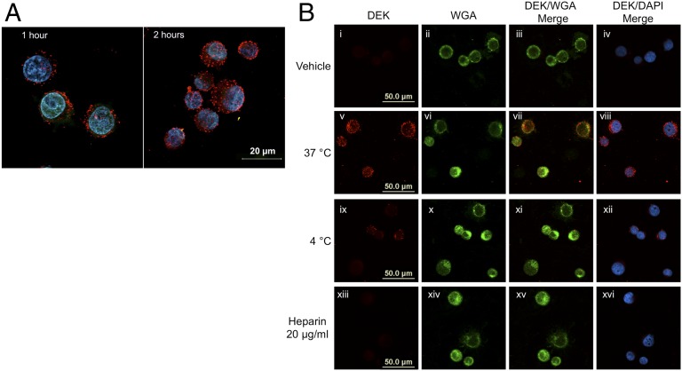
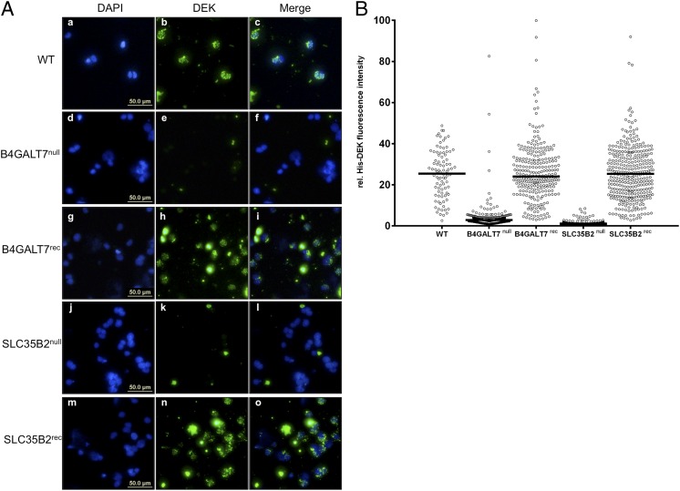
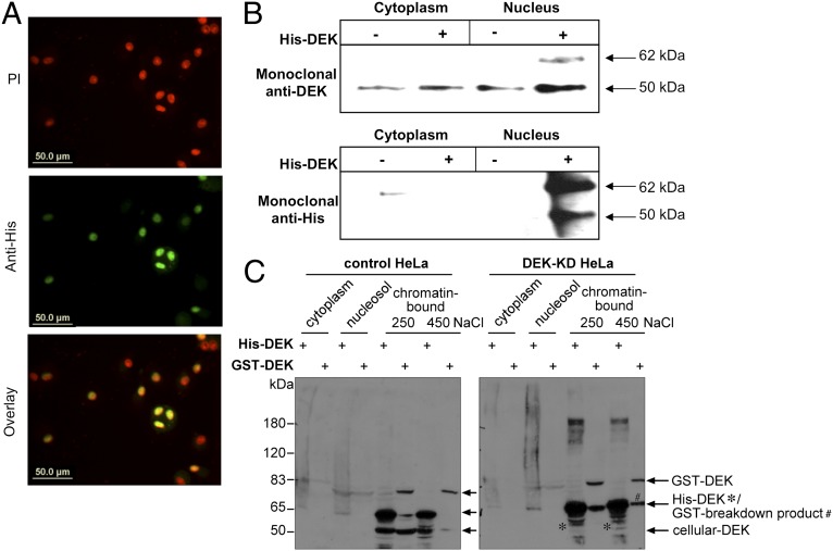
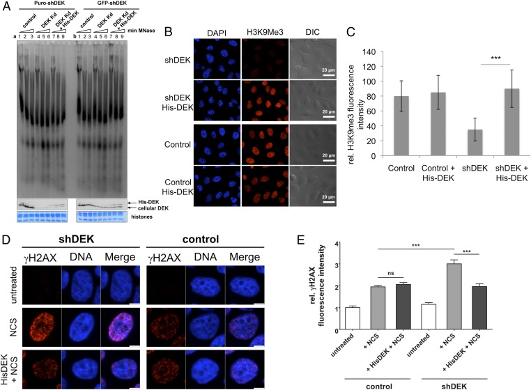
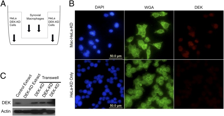
DEK is a biochemically distinct, conserved nonhistone protein that is vital to global heterochromatin integrity. In addition, DEK can be secreted and function as a chemotactic, proinflammatory factor. Here we show that exogenous DEK can penetrate cells, translocate to the nucleus, and there carry out its endogenous nuclear functions. Strikingly, adjacent cells can take up DEK secreted from synovial macrophages. DEK internalization is a heparan sulfate-dependent process, and cellular uptake of DEK into DEK knockdown cells corrects global heterochromatin depletion and DNA repair deficits, the phenotypic aberrations characteristic of these cells. These findings thus unify the extracellular and intracellular activities of DEK, and suggest that this paracrine loop involving DEK plays a role in chromatin biology.


Conflict of interest statement

The authors declare no conflict of interest.

Figures

Fig. 1.
Fig. 1.
DEK is taken up by cells in an energy-dependent process that requires surface HSPGs. (A) DEK-KD HeLa S3 cells were incubated with His-tagged recombinant DEK (20 µg/ 1 × 10⁷ cells) for 1 or 2 h and stained with DAPI (blue) and a monoclonal DEK antibody (red). (B) HeLa DEK-KD cells were incubated with recombinant His-DEK or vehicle for 2 h under varying conditions and stained with WGA (green), a membrane-specific stain, a monoclonal DEK antibody (red), and DAPI (blue) to visualize the nuclei. (i–iv) Cells incubated with vehicle (buffer) alone. (v–viii) Cells incubated with recombinant His-DEK at 37 °C. (ix–xii) Cells incubated with recombinant His-DEK at 4 °C. (xiii–xvi) Cells pretreated with 20 μg/mL soluble heparin and then incubated with recombinant His-DEK and 20 μg/mL soluble heparin at 37 °C. Images were obtained by confocal microscopy.
Fig. 2.
Fig. 2.
Cells defective in essential enzymes required for HSPG synthesis or positioning exhibit significantly reduced DEK uptake. (A) WT or mutant HAP1 cells incubated with recombinant His-DEK at 37 °C for 2 h and stained with DAPI (blue) or a monoclonal DEK antibody (green). (a–c) WT HAP1 cells. (d–f) HAP1 cells deficient in B4GALT7. (g–i) HAP1 B4GALT7null cells reconstituted with functional B4GALT7. (j–l) HAP1 cells deficient in SLC35B2. (m–o) HAP1 SLC35B2null cells reconstituted with functional SLC35B2. (B) Graphical representation of His-DEK–specific intensities in the nucleus. Multiple micrographs, as shown in A, were subjected to an automated intensity measurement workflow using the KNIME image processing software. More details are shown in Fig. 4 and Figs. S3 and S4. Segmentation of nuclei was carried out in the DAPI channel, followed by measurement of the His-DEK–specific intensities within the assigned segments using the FITC channel. Signals originating from cell clumps, debris, dead cells, or background signals owing to nonspecific extracellular aggregation of His-DEK were excluded from the measurements. Median values for the relative (rel.) His-DEK fluorescence intensity of all samples in a given group are indicated by the horizontal lines in the scatter dot plot.
Fig. 3.
Fig. 3.
DEK taken up by cells migrates to chromatin. (A) Twelve-day MDMs in 40% human serum were treated overnight with 15 µg of recombinant His-tagged DEK. Cells were stained for DEK with a monoclonal anti-His antibody, and nuclei were stained with propidium iodide (PI). (B) Immunoprecipitation of DEK from cytoplasmic and nuclear fractions of cells treated as in A with a rabbit polyclonal anti-His antibody. The resulting samples were analyzed on an SDS gel and immunoblotted with anti-DEK or anti-His monoclonal antibodies. (C) Control or HeLa DEK-KD cells were incubated with recombinant His-DEK or GST-DEK for 24 h and fractionated into cytosolic, nucleosolic, and chromatin-bound fractions. Proteins from each fraction were precipitated, resolved by SDS/PAGE, and blotted for DEK using a polyclonal antibody.
Fig. 4.
Fig. 4.
Exogenous DEK is bioactive on uptake into cells. Control or HeLa DEK-KD cells were incubated with recombinant His-DEK or buffer alone for 48 h and then subjected to functional analysis. (A) Cellular nuclei from control or HeLa DEK-KD, selected either by puromycin (a) or by FACS for GFP expression (b), were isolated, adjusted to 500 μg DNA/mL, and subjected to micrococcal nuclease treatment (5 U/50 μg DNA) of increasing duration (1 min in lanes 1, 4, and 7; 2 min in lanes 2, 5, and 8; 4 min in lanes 3, 6, and 9). The DNA fragments thus produced were analyzed directly by agarose gel electrophoresis and ethidium bromide staining. Equal aliquots of the individual samples were further analyzed by immunoblotting using a DEK-specific antibody, confirming DEK knockdown (a and b, lanes 4–6) and the presence of His-DEK (a and b, lanes 7–9). Protein staining of core histones served as a loading control (histones). (B) Control or HeLa-KD (shDEK) cells were fixed and stained with DAPI (blue) or an antibody that recognizes the H3K9Me3 heterochromatin mark (red) in the presence or absence of exogenously added His-DEK. Shown are representative magnifications of tile scans of three biological replicates (Fig. S4). DIC, differential interference contrast. (C) H3K9Me3 intensities in the individual samples were analyzed using the KNIME image processing software and a newly developed workflow (Figs. S3 and S4). One out of three replicates is shown. On average, 1,500–2,000 cells were analyzed per sample. Datasets were compared using the unpaired Student t test. ***P = 0.0004. (D) Control or HeLa-KD (shDEK) cells were treated with NCS for 15 min to induce DNA double-strand breaks, fixed, and stained with DAPI (blue) or an antibody specific for γH2AX (red), a marker for DNA double-stranded breaks. (E) Graphical representation of γH2AX-specific fluorescence intensity in the nucleus. The fluorescence intensity of γH2AX-positive nuclei was significantly reduced in NCS-treated HeLa-KD cells incubated with exogenous His-DEK. At least 150 nuclei were analyzed per experimental condition. Datasets were compared using the unpaired Student t test. ***P = 0.0001.
Fig. 5.
Fig. 5.
Secreted DEK is taken up by adjacent cells. (A) Diagram of the Transwell apparatus used in these experiments. Synovial macrophages were seeded on the top layer of the Transwell apparatus with 0.4-μm pores. HeLa-KD cells were then seeded on the bottom layer 2 d later. Cells were incubated for 3 d, and HeLa extracts were analyzed by immunofluorescence imaging and immunoblot analysis. (B) Immunofluorescence images of HeLa-KD cells with (Mac-HeLa-KD) or without (HeLa-KD only) synovial macrophages on the top layer of the Transwell apparatus. Cells were stained with DAPI (blue), membrane-specific WGA (green), and a polyclonal DEK antibody (red). (C) Western blot of whole cell extracts from control HeLa cells, HeLa DEK-KD cells without synovial macrophages on the top layer of the Transwell apparatus, and HeLa DEK-KD cells with synovial macrophages on the top layer of the Transwell (last three lanes, representing three biological replicates).

References

    1. von Lindern M, Poustka A, Lerach H, Grosveld G. The (6;9) chromosome translocation, associated with a specific subtype of acute nonlymphocytic leukemia, leads to aberrant transcription of a target gene on 9q34. Mol Cell Biol. 1990;10(8):4016–4026. - PMC - PubMed
    1. von Lindern M, et al. The translocation (6;9), associated with a specific subtype of acute myeloid leukemia, results in the fusion of two genes, dek and can, and the expression of a chimeric, leukemia-specific dek-can mRNA. Mol Cell Biol. 1992;12(4):1687–1697. - PMC - PubMed
    1. Kappes F, et al. The DEK oncoprotein is a Su(var) that is essential to heterochromatin integrity. Genes Dev. 2011;25(7):673–678. - PMC - PubMed
    1. McGarvey T, et al. The acute myeloid leukemia-associated protein, DEK, forms a splicing-dependent interaction with exon-product complexes. J Cell Biol. 2000;150(2):309–320. - PMC - PubMed
    1. Soares LM, Zanier K, Mackereth C, Sattler M, Valcárcel J. Intron removal requires proofreading of U2AF/3′ splice site recognition by DEK. Science. 2006;312(5782):1961–1965. - PubMed

Publication types