. 2013 Apr 23;110(17):6859-64.
doi: 10.1073/pnas.1219871110. Epub 2013 Apr 8.

Newly characterized Golgi-localized family of proteins is involved in calcium and pH homeostasis in yeast and human cells

Affiliations

Newly characterized Golgi-localized family of proteins is involved in calcium and pH homeostasis in yeast and human cells

Didier Demaegd et al. Proc Natl Acad Sci U S A. .

Abstract

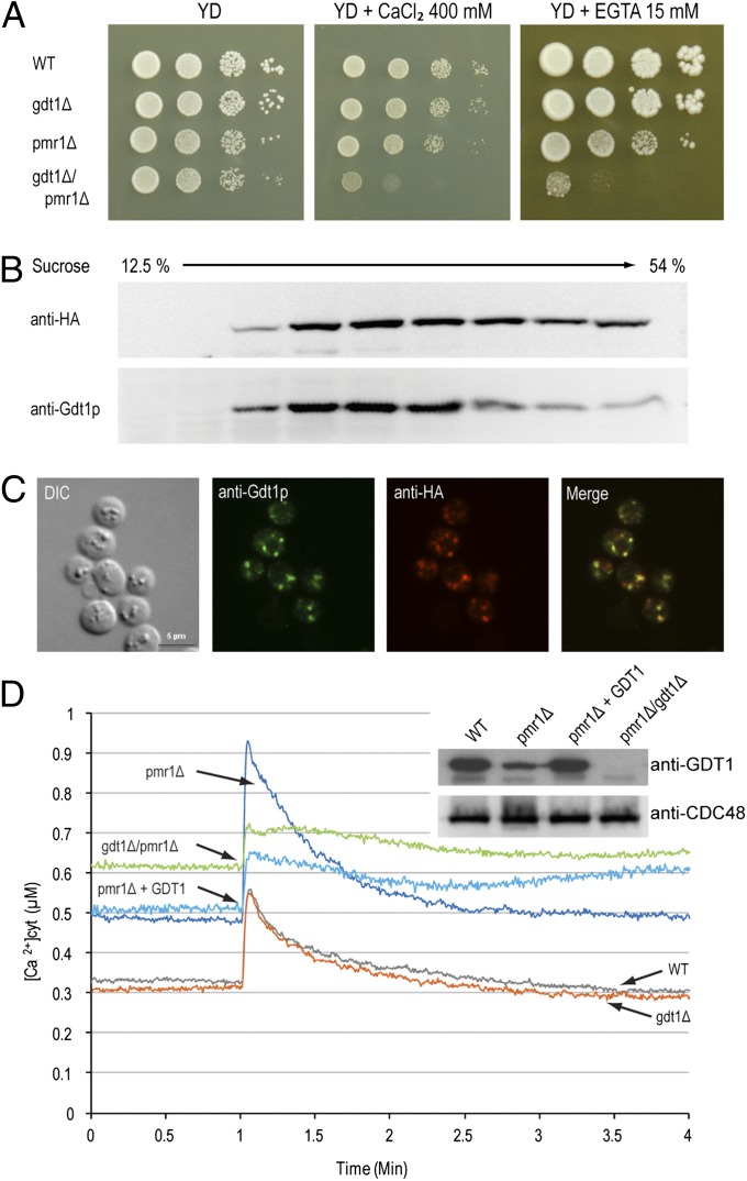
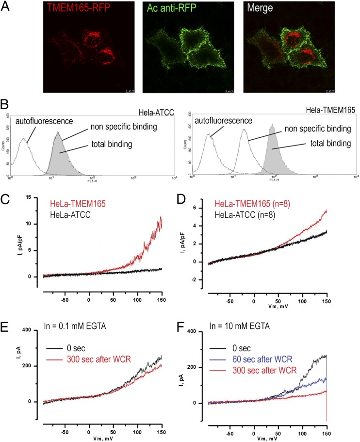
Defects in the human protein TMEM165 are known to cause a subtype of Congenital Disorders of Glycosylation. Transmembrane protein 165 (TMEM165) belongs to an uncharacterized family of membrane proteins called Uncharacterized Protein Family 0016, which are well conserved throughout evolution and share characteristics reminiscent of the cation/Ca(2+) exchanger superfamily. Gcr1 dependent translation factor 1 (Gdt1p), the budding yeast member of this family, contributes to Ca(2+) homeostasis via an uncharacterized Ca(2+) transport pathway localized in the Golgi apparatus. The gdt1Δ mutant was found to be sensitive to high concentrations of Ca(2+), and interestingly, this sensitivity was suppressed by expression of TMEM165, the human ortholog of Gdt1p, indicating conservation of function among the members of this family. Patch-clamp analyses on human cells indicated that TMEM165 expression is linked to Ca(2+) ion transport. Furthermore, defects in TMEM165 affected both Ca(2+) and pH homeostasis. Based on these results, we propose that Gdt1p and TMEM165 could be members of a unique family of Golgi-localized Ca(2+)/H(+) antiporters and that modification of the Golgi Ca(2+) and pH balance could explain the glycosylation defects observed in TMEM165-deficient patients.


Conflict of interest statement

The authors declare no conflict of interest.

Figures

Fig. 1.
Fig. 1.
GDT1 genetically interacts with PMR1. (A) The different mutants were precultured in YD medium to an OD600 of 0.3, then serial 10-fold dilutions were dropped onto solid YD medium alone or supplemented with 400 mM CaCl2 or 15 mM EGTA and the plates incubated at 28 °C for 4–6 d. Colocalization of Gdt1p and HA-Pmr1p as shown by subcellular fractionation (B) or immunostaining (C). (B) The fractions collected from the top of a discontinuous 10-step sucrose gradient were analyzed by Western blotting using antibodies against HA-Pmr1p (anti-HA) or Gdt1p. (C) Gdt1p and HA-Pmr1p in yeast were labeled using rabbit or rat primary antibodies, respectively, and fluorescein-conjugated anti-rabbit IgG and Alexa Fluor 546–conjugated anti-rat IgG antibodies. Differential interference contrast (DIC) and merged (merge) pictures are also presented. (Scale bar, 5 µm.) (D) The different strains expressing apo-aequorin from a plasmid were incubated overnight in minimal medium containing coelenterazine to reconstitute the holoenzyme. When the OD600 reached 3.0, 200 µl of each culture was transferred to a luminometric tube and the baseline luminescence monitored for 60 s, then CaCl2 was injected to a final concentration of 133 mM and the signal monitored for 3 min. The [Ca2+]cyt values were derived from luminometric units using the equation from Allen et al. (1977) (37). All displayed results are representative of those seen with at least three replicates. (Inset) Gdt1p and Cdc48p (loading control) levels in the different mutants measured by Western blotting of total extracts using anti-Gdt1p or anti-Cdc48p antibody. “pmr1Δ + GDT1” corresponds to the pmr1Δ mutant overexpressing GDT1.
Fig. 2.
Fig. 2.
Ionic movements are monitored across the plasma membrane when TMEM165 is overexpressed. (A) Living cells stably expressing TMEM165-RFP were incubated for 10 min at 18 °C with rabbit anti-RFP antibody and then were fixed and immunostained with Alexa Fluor 488–conjugated anti-rabbit IgG antibodies. (B) Presence of TMEM165 at the plasma membrane of HeLa cells stably expressing TMEM165-RFP investigated by flow cytometry. As described in Materials and Methods, fixed cells were incubated with rabbit anti-RFP antibodies and stained with Alexa Fluor 488–conjugated anti-rabbit IgG antibody. The plots show the green fluorescence (FL1-H) of wild-type HeLa cells (Left) and TMEM165-RFP–expressing HeLa cells (Right). Each plot shows the fluorescence of cells incubated without antibodies (autofluorescence; black line on left), only with Alexa Fluor 488–conjugated anti-rabbit IgG antibody (nonspecific binding; black line on right), or with both anti-RFP rabbit antibodies and Alexa Fluor 488–conjugated anti-rabbit antibody (total binding; gray shaded area). (C) Whole-cell membrane currents were measured in nontransfected HeLa cells (HeLa–ATCC) and HeLa cells overexpressing TMEM165. (D) Averaged membrane currents for n = 8. (E and F) Membrane currents in HeLa cells overexpressing TMEM165 in the presence of 0.1 mM EGTA (E) or 10 mM EGTA (F) (n = 4). WCR, whole-cell recording.
Fig. 3.
Fig. 3.
Lysosomal pH homeostasis is disturbed in TMEM165-deficient patients’ cells. (A) Live cell fluorescent images of lysosomal pH using LysoSensor green DND189 in fibroblasts from a control (C), five TMEM165-deficient patients (P1 to P5), and two V0-ATPase–deficient patients (P6 and P7). (B) Quantification of the fluorescence intensities observed in A. Error bars indicate mean ± 95% confidence interval (n = 3, around 10 cells per experiments). Data were analyzed by one-way ANOVA followed by Student post hoc test to identify mean differences between treatment and positive control. P values less than 0.05, 0.01, or 0.001 were indicated by *, **, or ***, respectively. Statistical analyses were performed using the software JMP 10 (SAS Institute). (C) Control and patients’ fibroblasts were incubated with LysoSensor green DND189, harvested, and analyzed by flow cytometry. Each histogram shows the autofluorescence (black line on the left) and the fluorescence (FL1-H) for the control cells (black line on the right) and one of the TMEM165-deficient patients’ cells (filled gray area). (D) Quantification of fluorescence intensities. The results were expressed as the percentage of the specific staining of the control cells—that is, the difference between the total and nonspecific binding peaks (autofluorescence). (E and F) HeLa cells were transfected with nontargeting siRNA (control) or siRNA targeted against TMEM165 (siRNA TMEM165); then, 7 d later, indirect immunofluorescence (E) and Western blotting analysis (F) were performed using anti-TMEM165 antibodies. (G) Live cell fluorescent images of the lysosomal pH using LysoSensor Green DND189 in control HeLa cells and TMEM165 siRNA knock-down HeLa cells. The results shown are representative of multiple cells assessed in independent experiments. (H) Quantification of the green fluorescence intensity (error bars indicate mean ± SEM, n = 3, >15 cells per experiments).

References

    1. Foulquier F, et al. TMEM165 deficiency causes a congenital disorder of glycosylation. Am J Hum Genet. 2012;91(1):15–26. - PMC - PubMed
    1. Wu X, et al. Mutation of the COG complex subunit gene COG7 causes a lethal congenital disorder. Nat Med. 2004;10(5):518–523. - PubMed
    1. Reynders E, et al. Golgi function and dysfunction in the first COG4-deficient CDG type II patient. Hum Mol Genet. 2009;18(17):3244–3256. - PMC - PubMed
    1. Foulquier F, et al. A new inborn error of glycosylation due to a Cog8 deficiency reveals a critical role for the Cog1-Cog8 interaction in COG complex formation. Hum Mol Genet. 2007;16(7):717–730. - PubMed
    1. Reynders E, Foulquier F, Annaert W, Matthijs G. How Golgi glycosylation meets and needs trafficking: The case of the COG complex. Glycobiology. 2011;21(7):853–863. - PubMed

Publication types

MeSH terms