

Intraductal papilloma of the breast in association with preoncogenic gene of breast cancer
- PMID: 23569749
- PMCID: PMC3609165
- DOI: 10.1016/S2221-1691(11)60017-8
Intraductal papilloma of the breast in association with preoncogenic gene of breast cancer
Abstract
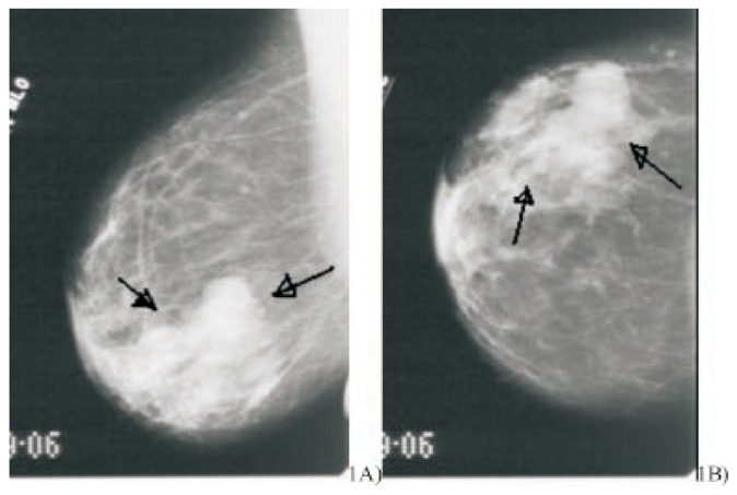
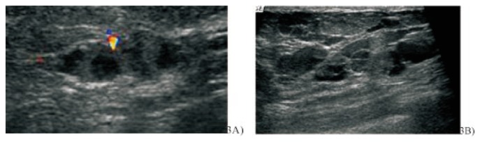
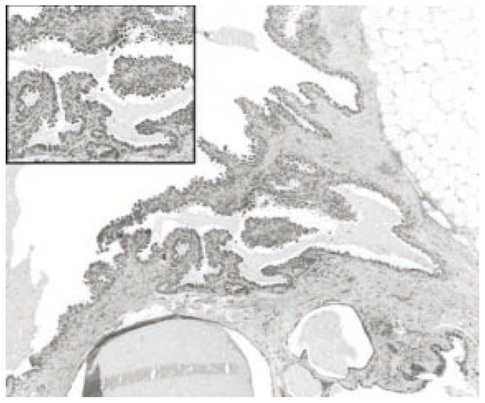
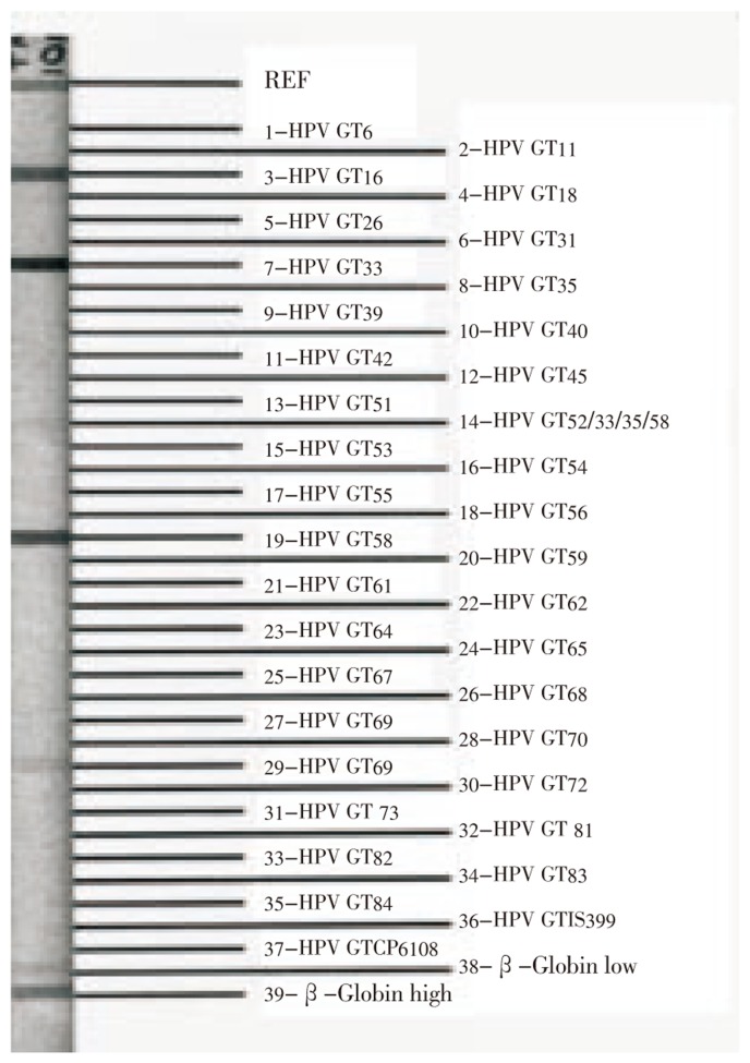
We reported a case of an African American woman who went to the hospital with palpable right breast lump with bloody nipple discharge at University of Texas Medical Branch at Galveston. The modalities of breast imagings included mammography and ultrasonography. The method used for viral identification was Linear Array HPV genotyping test. Intraductal papilloma revealed as high density tubular or rounded lobular masses with partially circumscribed, obscured margins and clustered punctate microcalcifications on mammograms. Ultrasound showed as intraductal masses with dilated ducts. The core biopsy demonstrated duct filled with papillary lesion and post excision revealed intraductal papilloma. HPV DNA types 16, 33, 58 and 71 were detected after use of Linear Array HPV genotyping test.
Keywords: Breast cancer; Human papilloma virus; Mammography; Preoncogenic gene; Ultrasound.
Conflict of interest statement
Figures

Similar articles
-
A retrospective observational study of intraductal breast papilloma and its coexisting lesions: A real-world experience.Cancer Med. 2020 Oct;9(20):7751-7762. doi: 10.1002/cam4.3308. Epub 2020 Aug 21. Cancer Med. 2020. PMID: 32822113 Free PMC article.
-
Is human papillomavirus associated with breast cancer or papilloma presenting with pathologic nipple discharge?Cancer Treat Res Commun. 2019;19:100122. doi: 10.1016/j.ctarc.2019.100122. Epub 2019 Feb 6. Cancer Treat Res Commun. 2019. PMID: 30785026
-
Radiologic and Pathologic Findings of Axillary Intraductal Papilloma Arising in Accessory Breast Tissue: A Case Report and Literature Review.Curr Med Imaging. 2022;18(14):1526-1528. doi: 10.2174/1573405618666220511193557. Curr Med Imaging. 2022. PMID: 35546773 Review.
-
[Mammography and magnetic resonance imaging for diagnosis of the intraductal papilloma of the breast].Nan Fang Yi Ke Da Xue Xue Bao. 2009 Aug;29(8):1643-6. Nan Fang Yi Ke Da Xue Xue Bao. 2009. PMID: 19726318 Chinese.
-
A case of giant complicated intraductal papilloma of breast on MRI and literature review.Cancer Rep (Hoboken). 2018 Dec;1(4):e1136. doi: 10.1002/cnr2.1136. Epub 2018 Sep 24. Cancer Rep (Hoboken). 2018. PMID: 32729233 Free PMC article. Review.
Cited by
-
Human papillomaviruses and breast cancer: A systematic review and meta‑analysis.Oncol Lett. 2023 Dec 22;27(2):75. doi: 10.3892/ol.2023.14208. eCollection 2024 Feb. Oncol Lett. 2023. PMID: 38192655 Free PMC article.
-
Breast Disorders in Adolescence: A Review of the Literature.Breast Care (Basel). 2021 Apr;16(2):149-155. doi: 10.1159/000511924. Epub 2020 Nov 30. Breast Care (Basel). 2021. PMID: 34012369 Free PMC article. Review.
References
-
- American Cancer Society . Cancer facts & figures 2007. Atlanta: American Cancer Society; 2007.
-
- American Cancer Society . What are the key statistics for breast cancer? Atlanta: American Cancer Society; 2006.
-
- Longnecker MP, Bernstein L, Paganini-Hill A, Enger SM, Ross RK. Risk factors for in situ breast cancer. Cancer Epidemiol Biomarkers Prev. 1996;5:961–965. - PubMed
-
- Trentham-Dietz A, Newcomb PA, Storer BE, Remington PL. Riskfactors for carcinoma in situ of the breast. Cancer Epidemiol Biomarkers Prev. 2000;9:697–703. - PubMed
-
- Band PR, Nhu DL, Fang R, Deschamps M. Carcinogenic and endocrine disrupting effects of cigarette smoke and risk of breast cancer. Lancet. 2002;360:1044–1049. - PubMed
Publication types
MeSH terms
LinkOut - more resources
Full Text Sources
Medical
