

A case report of pulmonary coinfection of Strongyloides stercoralis and Pneumocystis jiroveci
- PMID: 23569788
- PMCID: PMC3614232
- DOI: 10.1016/S2221-1691(11)60056-7
A case report of pulmonary coinfection of Strongyloides stercoralis and Pneumocystis jiroveci
Abstract
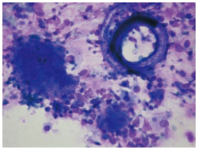
A case of pulmonary coinfection by Strongyloides stercoralis and Pneumocystis jiroveci has been detected in an AIDS patient treated in the Respiratory Intensive Care Unit of the Muñiz Hospital. At diagnosis, the patient presented cough with mucopurulent expectoration, dyspnea, fever, bilateral pulmonary infiltrates on the chest X-ray, negative bacilloscopy for acid fast bacteria and a CD4(+) T lymphocytes count of 52 cells/µL. The microbiological diagnosis was achieved by microscopic observation of the respiratory secretions obtained by bronchoalveolar lavage, while the wet mount examination revealed rhabditiform and filariform larvae of the nematode and foamy exudates, pathognomonic of the pulmonary pneumocystosis. It was the unique case of this association among about 3 000 samples performed in our laboratory in the last 10 years and diagnosed by microscopy. Other complementary stains (a rapid modification of Grocott, Kinyoun and Giemsa) were applied to the smears after the diagnosis of mycotic and parasitary infections achieved by fresh microscopy. Both physicians and microbiologists should take into account the possible coexistence of respiratory pathogens in immunocompromised patients, such as those with AIDS.
Keywords: AIDS; Bronchoalveolar lavage; Pneumocystis jiroveci; Pulmonary coinfection; Pulmonary pneumocystosis; Strongyloides stercoralis; Wet mout examination.
Conflict of interest statement
Figures
References
-
- Wazir JF, Ansari NA. Pneumocystis carinii infection: update and review. Arch Pathol Lab Med. 2004;128:1023–1027. - PubMed
-
- Mootsikapun P, Chetchotisakd P, Intarapoka B. Pulmonary infections in HIV infected patients. J Med Assoc Thai. 1996;79:477–485. - PubMed
-
- Bava AJ, Bellegarde JE. La coloracion de Kinyoun aplicada a muestras fecales de pacientes con SIDA. Rev Argent Infectol. 2002;15:19–21.
-
- Bava AJ. Coloración rápida para la identificación de quistes de Pneumocystis carinii en materiales respiratorios. Acta Bioquím Clín Latinoam. 2003;37:189–192.
Publication types
MeSH terms
LinkOut - more resources
Full Text Sources
Research Materials
