Comparative Study
. 2013 Jun 6;121(23):4740-8.
doi: 10.1182/blood-2013-01-476333. Epub 2013 Apr 9.

CXCL13 plus interleukin 10 is highly specific for the diagnosis of CNS lymphoma

Affiliations
Comparative Study

CXCL13 plus interleukin 10 is highly specific for the diagnosis of CNS lymphoma

James L Rubenstein et al. Blood. .

Abstract

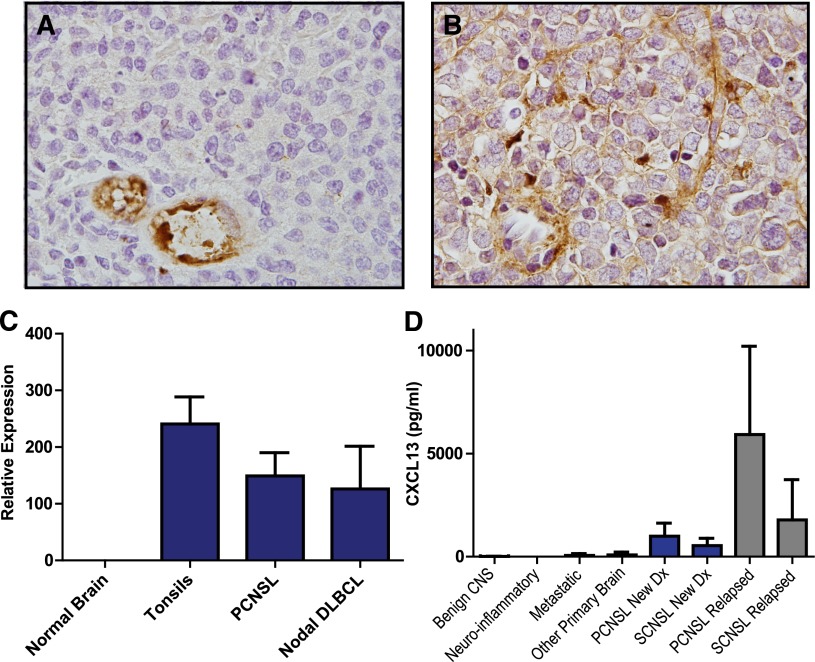
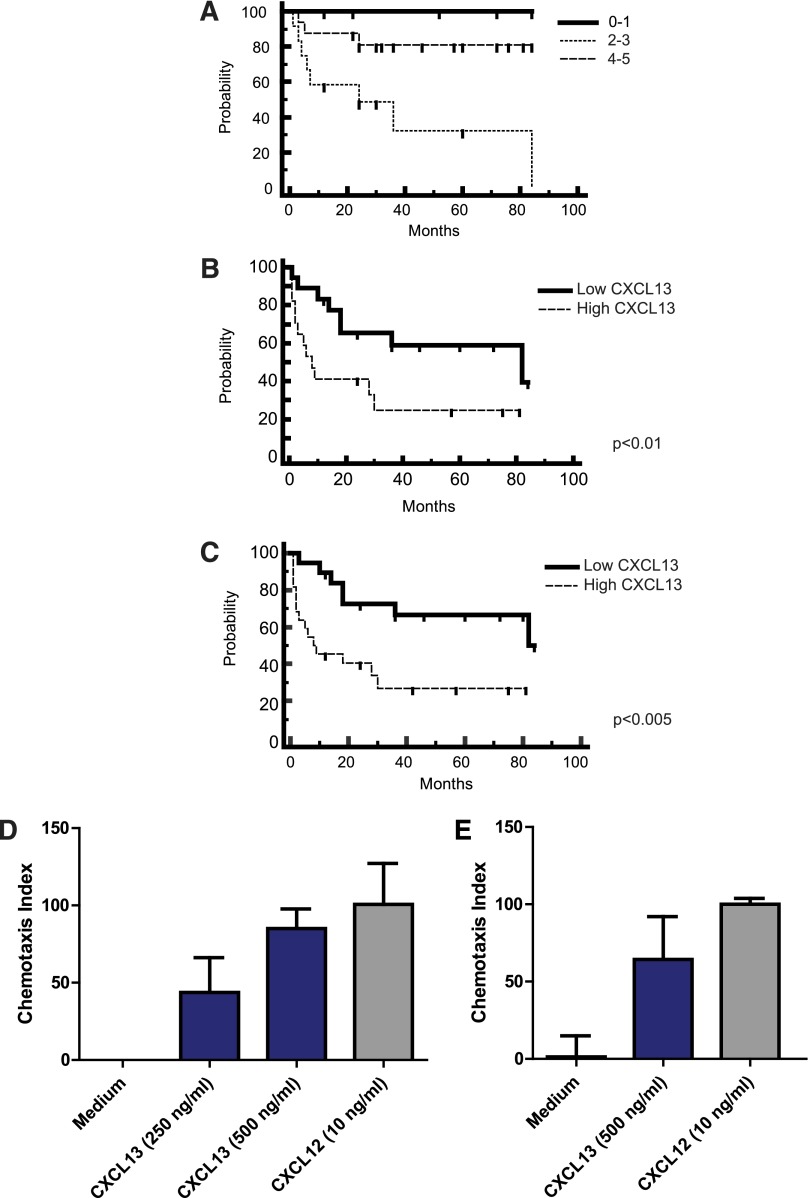
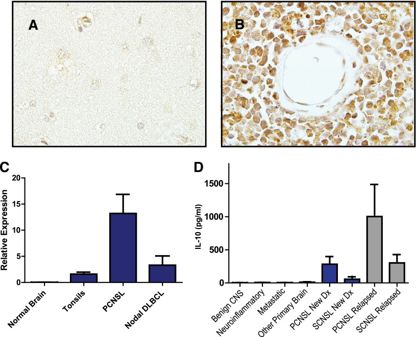
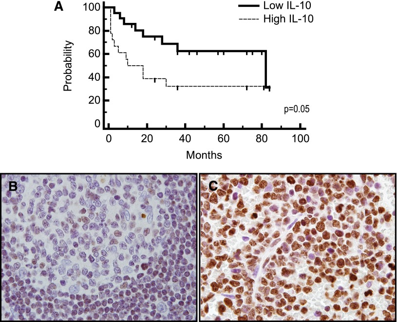
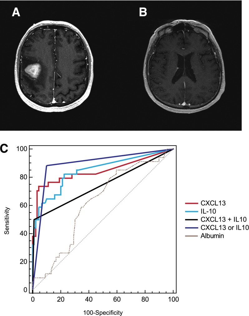
Establishing the diagnosis of focal brain lesions in patients with unexplained neurologic symptoms represents a challenge. The goal of this study is to provide evidence supporting functional roles for CXC chemokine ligand (CXCL)13 and interleukin (IL)-10 in central nervous system (CNS) lymphomas and to evaluate the utility of each as prognostic and diagnostic biomarkers. We demonstrate for the first time that elevated CXCL13 concentration in cerebrospinal fluid (CSF) is prognostic and that CXCL13 and CXCL12 mediate chemotaxis of lymphoma cells isolated from CNS lymphoma lesions. Expression of the activated form of Janus kinase 1 supported a role for IL-10 in prosurvival signaling. We determined the concentration of CXCL13 and IL-10 in CSF of CNS lymphoma patients and control cohorts including inflammatory and degenerative neurologic disease in a multicenter study involving 220 patients. Bivariate elevated CXCL13 plus IL-10 was 99.3% specific for primary and secondary CNS lymphoma, with sensitivity significantly greater than reference standard CSF tests. These results identify CXCL13 and IL-10 as potentially important biomarkers of CNS lymphoma that merit further evaluation and support incorporation of CXCL13 and IL-10 into diagnostic algorithms for the workup of focal brain lesions in which lymphoma is a consideration.


Figures

Figure 1
Figure 1
CXCL13 expression in CNS lymphomas. Expression of CXCL13 in a diagnostic specimen of de novo PCNSL (A) and in relapsed SCNSL (B), as demonstrated by immunohistochemistry (×1000). CXCL13 expression is most evident in stromal elements including tumor blood vessels. (C) Quantitative RT-PCR demonstrates markedly increased expression of CXCL13 in diagnostic specimens of PCNSL (N = 23). CXCL13 transcript levels were similar in PCNSL compared with reactive pharyngeal tonsils (N = 2) and nodal DLBCL (N = 9) but barely detectable in specimens of normal brain (N = 3 cases). (D) CXCL13 protein is markedly increased in CSF in association with both PCNSL and SCNSL compared with the vast majority of neuro-inflammatory conditions and other brain tumors. The highest CSF concentration of CXCL13 was detected in association with relapsed CNS non-Hodgkin lymphoma.
Figure 2
Figure 2
CXCL13 is prognostic and mediates chemotaxis of lymphoma cells isolated from CNS lymphoma lesions. (A) Survival for the cohort of PCNSL patients by International Extranodal Lymphoma Study Group score (N = 33 total patients), demonstrating representation of the prognostic subgroups. Kaplan-Meier analysis; y-axis indicates percent overall survival (OS). The International Extranodal Lymphoma Study Group prognostic score is based on 5 variables: age >60, Performance Status >1, elevated lactate dehydrogenase, elevated CSF protein, and involvement of deep regions in the brain. (B) Newly diagnosed patients with PCNSL (N = 34 patients) with elevated CXCL13 at diagnosis (>200 pg/mL, the median CSF concentration among all CNS lymphoma patients) exhibited shorter time to progression (TTP) after treatment with methorexate-based induction. P < .01 (hazard ratio = 2.96). The median TTP of PCNSL patients with low CSF CXCL13 (N = 16) is 82 months vs 7 months for patients who presented with elevated CSF CXCL13. (C) PCNSL and SCNSL (N = 41) with elevated CSF CXCL13 at diagnosis exhibit shorter PFS (defined as disease progression or death as a result from any cause). P < .005 (hazard ratio = 3.12). The median PFS of PCNSL and SCNSL patients with low CSF CXCL13 (N = 19) is 82 months vs 9 months for patients who presented with high CXCL13. The OS of PCNSL and SCNSL patients with low CSF CXCL13 at diagnosis is also longer than for patients with high CSF CXCL13 at diagnosis. P < .04 (hazard ratio = 3.03), although the median OS for both cohorts has not been reached (not shown). All patients were treated with a high-dose methotrexate-based induction regimen without whole brain irradiation consolidation. Patients with stage IV DLBCL with CNS involvement (C) received cyclophosphamide, vincristine, adriamycin, and prednisone instead of temozolomide, as described. (D) Directed chemotaxis of CNS lymphoma cells (isolated from a brain parenchymal lesion of relapsed CNS lymphoma) in response to chemokines CXCL13 and CXCL12. The y-axis depicts chemotaxis index (% compared with medium control). P < .05. (E) CXCL13- and CXCL12-mediated chemotaxis of meningeal lymphoma cells isolated from the CSF of a CNS lymphoma patient with refractory disease. P < .05. There was no evidence for synergy or additive effects when CXCL13 and CXCL12 were used in combination, and there was no chemotaxis in response to C3a anaphylatoxin or ephrin A4 peptides.
Figure 3
Figure 3
IL-10 expression in CNS lymphomas. Absent IL-10 expression in normal brain (A) with strong expression of IL-10 by lymphoma cells in PCNSL (B), as demonstrated by immunohistochemistry. There was weak or absent IL-10 expression by tumor vessels (×1000). (C) Quantitative RT-PCR demonstrates markedly increased expression of IL-10 in diagnostic specimens of PCNSL (N = 23) compared with reactive tonsils and specimens of normal brain. The average IL-10 transcriptional expression was also higher in PCNSL compared with 9 cases of nodal DLBCL, of which 7 were of germinal center B-cell phenotype. Notably, the normalized IL-10 transcript level, 12, in the 1 nodal activated B-cell-type DLBCL specimen was similar to the mean-normalized IL-10 expression in the PCNSL cases. (D) Mean CSF IL-10 protein is >70-fold higher in patients with both PCNSL and SCNSL compared with neuro-inflammatory conditions and other brain tumors (P < 2.3 × 10⁻⁵). The CSF concentration of IL-10 was highest in relapsed cases.
Figure 4
Figure 4
IL-10 is prognostic and may be associated with JAK-1 activation. (A) Patients with non-HIV–associated PCNSL and SCNSL with elevated concentration of IL-10 in CSF at diagnosis (N = 18) (above 45 pg/mL, the median concentration among all CNS lymphoma patients) exhibited significantly shorter TTP compared with patients with low CSF IL-10 (N = 21). P = .05 (hazard ratio = 2.33). The median TTP (as well as PFS) of PCNSL and SCNSL patients with low CSF IL-10 at diagnosis is 82 months vs 10 months for patients with elevated CSF IL-10. The median OS of CNS lymphoma patients with low CSF IL-10 at diagnosis was 84 months, whereas the median OS of patients with elevated CSF IL-10 at diagnosis has not been reached. P < .057 (hazard ratio = 3.6) (not shown). All patients were treated with a high-dose methotrexate-based induction regimen without whole brain irradiation consolidation. Patients with stage IV DLBCL with CNS involvement received cyclophosphamide, vincristine, adriamycin, and prednisone instead of temozolomide, as described. Germinal center B cells in reactive tonsils (B) exhibited weak to absent immunoreactivity for JAK-1 activation (phosphorylation of JAK-1 at tyrosine 1022), whereas strong intratumoral JAK-1 activation (>30% lymphoma cells) was detected in 70% of diagnostic specimens of PCNSL (N = 30) (C) (×1000).
Figure 5
Figure 5
IL-10 gene expression and CSF concentration correlate with disease course in a patient with recurrent CNS lymphoma. (A) A decline in CSF concentration of IL-10 correlates with initial cytological response followed by tumor progression in a representative patient with recurrent SCNSL who participated in a phase 1 trial of intraventricular rituximab plus methotrexate. (The result is representative of 6 consecutive trial patients analyzed.) (B) Cytological appearance of lymphoma cells in CSF at baseline and persistent disease at completion of intraventricular therapy (28 days). (C) Quantitative RT-PCR of RNA transcripts purified from B cells isolated from the CSF by flow cytometry demonstrated upregulated IL-10 expression in refractory CNS lymphoma after treatment with rituximab plus methotrexate. The increased relative expression of IL-10 transcripts by the meningeal lymphoma cells corresponded to increased concentration of IL-10 protein in CSF at day 28 (A). (D) In contrast, transcriptional expression of IL-10 receptor A decreased after therapy.
Figure 6
Figure 6
CXCL13 and IL-10 are highly specific for the diagnosis of PCNSL. (A-B) MRI features of PCNSL in 2 patients at diagnosis. (A) MRI depicts a homogeneously contrast-enhancing mass with vasogenic edema. At the time of diagnosis established by brain biopsy, the CSF contained CXCL13 concentration of 170 pg/mL and IL-10 concentration of 61 pg/mL. (B) Normal-appearing MRI of a patient with progressive neurologic symptoms who was aggressively treated with steroids before a diagnosis could be elicited. Four CSF collections and 1 brain biopsy were unrevealing, and the diagnosis of disseminated PCNSL was made at autopsy. The CSF collected and stored from this patient was later determined to contain CXCL13 concentration of 6236 pg/mL and IL-10 concentration of 76 pg/mL. (C) ROC analysis demonstrated that high concentrations of CXCL13 and IL-10 are each highly sensitive and specific for CNS lymphomas. A CSF concentration of CXCL13 >116 pg/mL is 71% sensitive and 94.9% specific for PCNSL (area under the curve [AUC], 0.841; 95% confidence interval [CI], 0.779-0.892). A CSF concentration of IL-10 >23 pg/mL is 63.9% sensitive and 94.1% specific for CNS involvement of lymphoma (AUC, 0.851; 95% CI, 0.789-0.901). Bivariate elevation of both CXCL13 plus IL-10 in CSF was 50% sensitive and 99.3% specific for CNS lymphoma (AUC, 0.746; 95% CI, 0.675-0.809). Elevation of either CXCL13 or IL-10 in CSF was 84.2% sensitive and 90.5% specific for the diagnosis of PCNSL (AUC, 0.874; 95% CI, 0.815-0.919). By contrast, the AUC of CSF albumin was 0.604 and significantly lower than CXCL13 or IL-10 (P < .0001).

References

    1. Josephson SA, Papanastassiou AM, Berger MS, et al. The diagnostic utility of brain biopsy procedures in patients with rapidly deteriorating neurological conditions or dementia. J Neurosurg. 2007;106(1):72–75. - PubMed
    1. Hall WA. The safety and efficacy of stereotactic biopsy for intracranial lesions. Cancer. 1998;82(9):1749–1755. - PubMed
    1. Javedan SP, Tamargo RJ. Diagnostic yield of brain biopsy in neurodegenerative disorders. Neurosurgery. 1997;41(4):823–828, discussion 828-830. - PubMed
    1. Ferreri AJ. How I treat primary CNS lymphoma. Blood. 2011;118(3):510–522. - PubMed
    1. Hormigo A, Abrey L, Heinemann MH, DeAngelis LM. Ocular presentation of primary central nervous system lymphoma: diagnosis and treatment. Br J Haematol. 2004;126(2):202–208. - PubMed

Publication types

MeSH terms