. 2013 May;21(5):461-73.
doi: 10.1016/j.jagp.2012.09.005. Epub 2013 Mar 15.

Short-term recognition memory correlates with regional CNS expression of microRNA-138 in mice

Affiliations

Short-term recognition memory correlates with regional CNS expression of microRNA-138 in mice

Erick T Tatro et al. Am J Geriatr Psychiatry. 2013 May.

Abstract

Objectives: We hypothesized that microRNA (miR) expression may be involved in memory function because it controls local protein translation at synapses and dendritic spines.

Design: Case-control animal study.

Methods: We assessed the miR repertoire in the hippocampus of young, 6-month-old (N = 18) mice compared with aged, 26-month-old (N = 23) mice and compared miR quantity to memory scores as determined by the novel object recognition task. We performed a histological brain regional analysis of miR-138, acyl protein thioesterase 1 (APT1) mRNA, and APT1 protein.

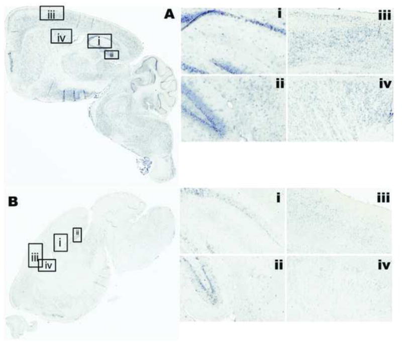
Results: We found that higher miR-138 expression in the mouse hippocampus is correlated with better memory performance. We also found that APT1 (a depalmytoylation enzyme expressed at dendritic spines whose translation is controlled by miR-138) mRNA is increased in the mouse hippocampal CA1 and dentate gyrus in aged mice compared with young mice, but not in mice with memory impairment. We found APT1 protein distribution to be lower in cells with high miR-138 expression.

Conclusions: These results suggest that increased miR-138 is associated with better memory and increased APT1 gene transcription occurs with aging. The role of miR-138 and APT1 protein function in memory and aging warrants further investigation.


Figures

Figure 1
Figure 1
Cognitive and behavioral features of the mouse cohort indicating lower object recognition as measured by DR in the aged group compared to the young (a) and no difference in distance traveled (b), center time (c), and sampling time (d) between impairment statuses. The line and heavy lines indicate mean and standard deviation. These data indicate lower memory performance in aging and variation in memory performance is not an artifact of variation in exploratory behavior.
Figure 2
Figure 2
MiR expression levels of 7 miRs from screen that were significantly different (p < 0.05) by two-sided one-way ANOVA comparing Young, Impaired-Aged, and Unimpaired-Aged groups. Illustrated are Log (Fold Change) values calibrated to average of young group, error bars indicate standard deviation (n = 4, each group) and experiment was performed in technical duplicate.
Figure 3
Figure 3
The distribution of miR-138 in situ hybridization in parasagittal section of mouse brain from a young unimpaired (a) and an aged impaired (b) mouse. Detailed regions are: CA1-CA2-CA3 of hippocampus (i), DG and subiculum of hippocampus (ii), motor cortex (iii), and caudate/putamen (iv). Fast red counterstain (insets) illustrate the presence of cells and nuclei in area of low to absent miR-138 in the aged-impaired specimen (b). For in situ hybridization positive control, probe for U6-small nuclear RNA (c), stains all nuclei, and negative control, scrambled LNA probe (d) illustrates any background coloration. MiR-138 in situ hybridization signal ranged from intense (a) to low (b) and was neuron-enriched in pyramidal neurons, small interneurons, and medium spiny neurons.
Figure 4
Figure 4
Values for miR-138 in situ hybridization signal in the neuroanatomic regions that were measured (IRn) in the dorsal hippocampal CA1 (a, c) and dorsal DG (b, d). The in situ hybridization signal was not different comparing by age; for CA1, t37.60 = −0.25, P = 0.4 (a) and for DG, t33.34 = −1.38, P = 0.08 (b). MiR-138 signal was significantly higher in CA1 of the unimpaired mice, CA1, t38.00 = 1.55 P = 0.06 (c), and DG, t37.72 = 1.82, P = 0.03 (d). Comparisons were made using one-sided Welch’s test, assuming unequal variances.
Figure 5
Figure 5
The distribution of APT1 mRNA in situ hybridization in parasagittal section of mouse brain from a young unimpaired (a) and an aged impaired (b) mouse. Detailed regions are: CA1-CA2-CA3 of hippocampus (i), DG and subiculum of hippocampus (ii), motor cortex (iii), and caudate/putamen (iv). In situ hybridization for APT1 mRNA was apparent in apical dendrites of pyramidal neurons. The signal was enriched in basal side of DG granular cells (ii). In the aged mouse, while expression was lower overall, APT1 mRNA appeared to be enriched near mossy fibers of CA1 (b–i).
Figure 6
Figure 6
Values for APT1 mRNA quantification separated by age (a–c) and impairment status (d–f). Quantification by qPCR from RNA isolated from frozen left-hippocampus homogenates, comparing by age (a) and impairment status (d), is illustrated with Log (fold-change) calibrated to average ΔCT value of the young group and using β-actin and GAPDH as endogenous controls. Values for in situ hybridization signal in the neuroanatomic regions that were measured (IRn) in the dorsal hippocampal CA1 (b, e) and dorsal DG (c, f). The line and heavy lines indicate mean and standard deviation. The qPCR signal was significantly different comparing by age, t30.69 = 4.21, P = 0.0002 (a) but not by impairment status, t36.88 = −0.42, P = 0.5 (d). APT1 in situ hybridization signal was not significantly different by age in CA1 (b), t37.00 = −0.08, P = 0.94, or in DG (c), t36.88 = −1.33, P = 0.09. Comparing by impairment status was not significantly different, t36.42 = 1.00, P = 0.48 for CA1 (e) and t26.61 = 0.17, P = 0.2 for DG (f).
Figure 7
Figure 7
The distribution of APT1 immunoreactivity in parasagittal section of mouse brain from a young unimpaired (a) and an aged impaired (b) mouse. Detailed regions are: CA1-CA2-CA3 of hippocampus (i), DG and subiculum of hippocampus (ii), motor cortex (iii), and caudate/putamen (iv). Immunoreactivity for APT1 was abundant in neuropil and lower in cell bodies.
Figure 8
Figure 8
Values for APT1 immunoreactivity in the neuroanatomic regions that were measured (IRn) in the dorsal hippocampal CA1 (a, c) and dorsal DG (b, d). The line and heavy lines indicate mean and standard deviation. The immunoreactivity signal was not different comparing by age, t35.95 =1.45, P = 0.16 in the CA1 (a) and t36.87 = −1.33 P = 0.19 in the DG (b). APT1 immunoreactivity was not significantly different by impairment status, t26.91 = −0.36, P = 0.72 for the CA1 (c) and t36.50 = 1.55, P = 0.13 for DG (d). Comparisons were made using two-sided Welch’s test, assuming unequal variances.
Figure 9
Figure 9
MiR-138 and APT1 in two serial sections each from a young/normal and aged/impaired specimen, showing 400X original magnification of dentate gyrus (a), pyramidal layer of the somato-motor cortex (b), caudate-putamen (c), CA3 of hippocampus (d), and thalamus (e). A schematic shows the location of the fields imaged fields (f), reproduced from Allen Brain Atlas (25). The sections labeled with in situ hybridization for miR-138 were developed with NBT/BCIP and counterstained using Fast Red (Left, all panels), while the sections immunolabeled for APT1 were developed with DAB and counterstained with haematoxylin (right, all panels). The 400X magnification shows APT1 immunoreactivity in some cell bodies and neuropil (right, all panels). In (a) the dentate gyrus, there was focal staining in oligodendrocytes of APT1 in cells lacking miR-138 (white arrowheads, top), and a lack of APT1 in the granular layer with abundant miR-138 (top). In the granular layer of the age/impaired specimen (bottom), there was some APT1 staining. In the pyramidal neurons of the frontal cortex (b), the young/normal specimen specimen had intense miR-138 and little in the aged/impaired mouse. The focal APT1 staining in oligodendrocytes was apparent in the FC as with the DG (white arrowheads). There was also a notable concentric staining in cell bodies (black arrowheads). In the CP, there was notably more miR-138 in the young/normal specimen compared to the aged/normal, with miR-138 in the medium spiny neurons, and there was less APT1 in these specific cells (white arrowhead, top). In cells with little miR-138, there was more APT1 (black arrowheads) in the neuropil and conversely, intense miR-138, there was less APT1 (white arrowheads, top). The APT1 staining in the CA3 granular layer seemed to mainly be concentric in cell bodies (black arrowheads, top and bottom). In this aged/impaired specimen, one APT1-positive oligodendrocyte was observed (white arrowhead, bottom) as in the DG and FC, but was not observed in this example of young/normal. In the thalamus (e), both miR-138 and APT1 were observed. The APT1 was both in the concentric, cell body staining (black arrowheads, top and bottom) and oligodendrocytes (white arrowheads, bottom). For miR-138, there was staining in many cell bodies of the young, normal specimen (top). Abbreviations: DG - dentate gyrus, FC - frontal cortex, CP - caudate-putamen, CA3 - CA3 field of the hippocampus, Thal - thalamus.

References

    1. Hernandez PJ, Abel T. The role of protein synthesis in memory consolidation: progress amid decades of debate. Neurobiol Learn Mem. 2008;89:293–311. - PMC - PubMed
    1. Ashraf SI, Kunes S. A trace of silence: memory and microRNA at the synapse. Curr Opin Neurobiol. 2006;16:535–539. - PubMed
    1. Bramham CR, Wells DG. Dendritic mRNA: transport, translation and function. Nat Rev Neurosci. 2007;8:776–789. - PubMed
    1. Martin KC, Barad M, Kandel ER. Local protein synthesis and its role in synapse-specific plasticity. Curr Opin Neurobiol. 2000;10:587–592. - PubMed
    1. Ostroff LE, Fiala JC, Allwardt B, et al. Polyribosomes redistribute from dendritic shafts into spines with enlarged synapses during LTP in developing rat hippocampal slices. Neuron. 2002;35:535–545. - PubMed

Publication types