

Generation and characterization of virus-free reprogrammed melanoma cells by the piggyBac transposon
- PMID: 23571855
- PMCID: PMC11824178
- DOI: 10.1007/s00432-013-1431-3
Generation and characterization of virus-free reprogrammed melanoma cells by the piggyBac transposon
Erratum in
- J Cancer Res Clin Oncol. 2013 Sep;139(9):1601. Fan, Yongna [added];Qin, Dingxin [added]; Xiaocui Bian, Xiaocui [added]
Abstract
Purpose: Reprogramming of cancer cells to stem cell-like state provides a promising tool for the study of cancer pathogenesis and drug screening. However, most instances of direct reprogramming have been achieved by forced co-expression of defined transcription factors using viral vectors. Retroviral transduction as well as the ectopic expression of reprogramming factors may alter the differentiation potential of reprogrammed cancer cells or induce malignant transformation. Therefore, generation of reprogrammed cancer cells via virus-free reprogramming strategy needs to be studied.
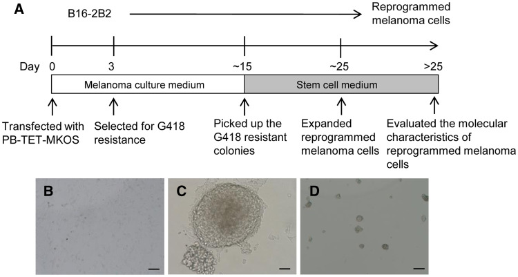
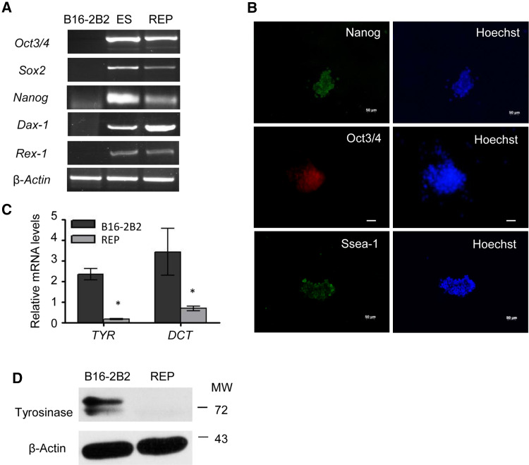
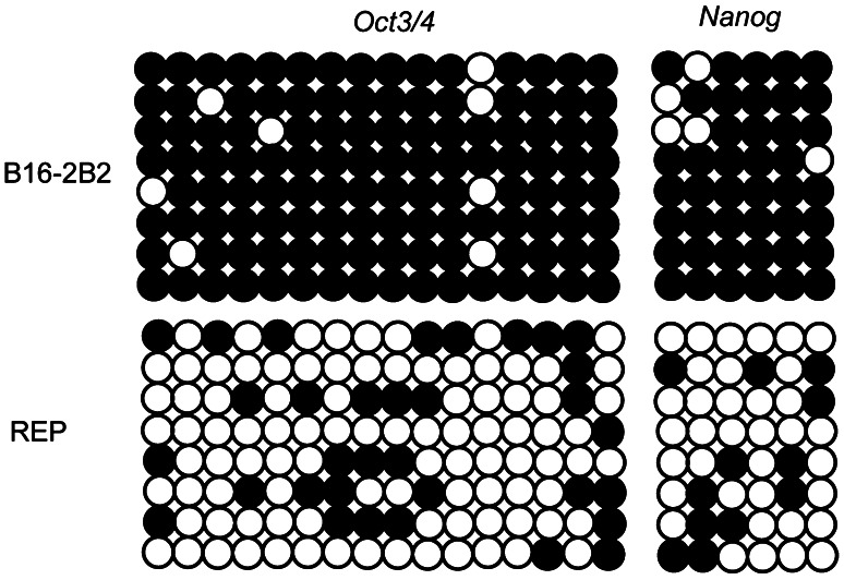
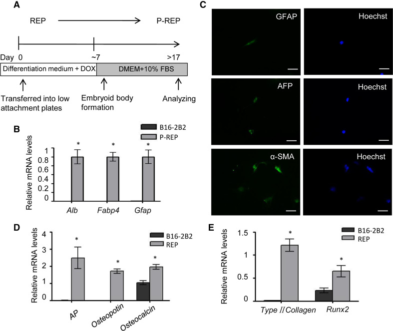
Methods: Melanoma cells were reprogrammed by co-expression of doxycycline-inducible Oct4, Sox2, Klf4, and c-Myc using the piggyBac (PB) transposon system. The expression level of genes was analyzed through RT-PCR, Western blot, and immunofluorescence. Epigenetic modification of genes was detected by bisulfite genomic sequencing. Post reprogrammed melanoma cells were generated through differentiation of reprogrammed melanoma cells. Sensitivity to chemotherapeutic agents and metastasis potential were investigated in post reprogrammed melanoma cells.
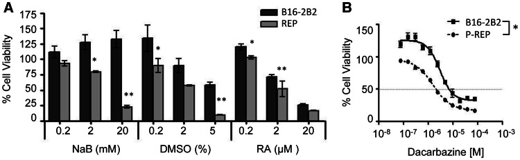
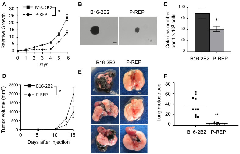
Results: The virus-free reprogrammed melanoma cells were positive for stem cell markers including Oct4, Nanog, and SSEA-1, and the promoters of Nanog and Oct4 were demethylated. Moreover, reprogrammed melanoma cells gained differentiation potential and higher sensitivity to differentiation-inducing drugs. Post reprogrammed melanoma cells showed lower proliferation rate and metastatic potential compared with the parental cells.
Conclusions: Our results indicate that PB transposon-based method is applicable to generate virus-free reprogrammed melanoma cells. These cells can differentiate into other lineages with loss of malignant phenotypes, which may provide a more suitable source for molding of cancer pathogenesis.
Conflict of interest statement
We declare that we have no conflict of interest.
Figures
Similar articles
-
Reprogramming factors induce proliferation and inhibit apoptosis of melanoma cells by changing the expression of particular genes.Mol Med Rep. 2019 Feb;19(2):967-973. doi: 10.3892/mmr.2018.9753. Epub 2018 Dec 12. Mol Med Rep. 2019. PMID: 30569122 Free PMC article.
-
OSKM Induce Extraembryonic Endoderm Stem Cells in Parallel to Induced Pluripotent Stem Cells.Stem Cell Reports. 2016 Apr 12;6(4):447-455. doi: 10.1016/j.stemcr.2016.02.003. Epub 2016 Mar 3. Stem Cell Reports. 2016. PMID: 26947975 Free PMC article.
-
Comparative analysis of mouse strains for in vivo induction of reprogramming factors.Cell Rep. 2025 Jul 22;44(7):115879. doi: 10.1016/j.celrep.2025.115879. Epub 2025 Jun 24. Cell Rep. 2025. PMID: 40560729
-
The Black Book of Psychotropic Dosing and Monitoring.Psychopharmacol Bull. 2024 Jul 8;54(3):8-59. Psychopharmacol Bull. 2024. PMID: 38993656 Free PMC article. Review.
-
Laboratory-based molecular test alternatives to RT-PCR for the diagnosis of SARS-CoV-2 infection.Cochrane Database Syst Rev. 2024 Oct 14;10(10):CD015618. doi: 10.1002/14651858.CD015618. Cochrane Database Syst Rev. 2024. PMID: 39400904
Cited by
-
Chinese Herbs Interfering with Cancer Reprogramming Metabolism.Evid Based Complement Alternat Med. 2016;2016:9282813. doi: 10.1155/2016/9282813. Epub 2016 May 5. Evid Based Complement Alternat Med. 2016. PMID: 27242914 Free PMC article. Review.
-
Application of induced pluripotency in cancer studies.Rep Pract Oncol Radiother. 2018 May-Jun;23(3):207-214. doi: 10.1016/j.rpor.2018.04.005. Epub 2018 Apr 24. Rep Pract Oncol Radiother. 2018. PMID: 29760595 Free PMC article. Review.
-
Sendai virus-mediated expression of reprogramming factors promotes plasticity of human neuroblastoma cells.Cancer Sci. 2015 Oct;106(10):1351-61. doi: 10.1111/cas.12746. Epub 2015 Aug 18. Cancer Sci. 2015. PMID: 26190440 Free PMC article.
References
-
- Degos L, Wang ZY (2001) All trans retinoic acid in acute promyelocytic leukemia. Oncogene 20:7140–7145 - PubMed
Publication types
MeSH terms
Substances
LinkOut - more resources
Full Text Sources
Other Literature Sources
Research Materials
