Review
. 2013 Nov-Dec;46(6):549-69.
doi: 10.1177/0022219413483174. Epub 2013 Apr 9.

Neurobiological underpinnings of math and reading learning disabilities

Affiliations
Review

Neurobiological underpinnings of math and reading learning disabilities

Sarit Ashkenazi et al. J Learn Disabil. 2013 Nov-Dec.

Abstract

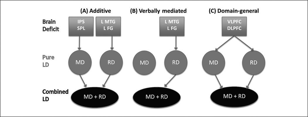
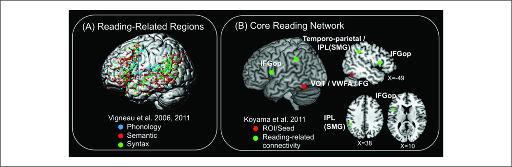
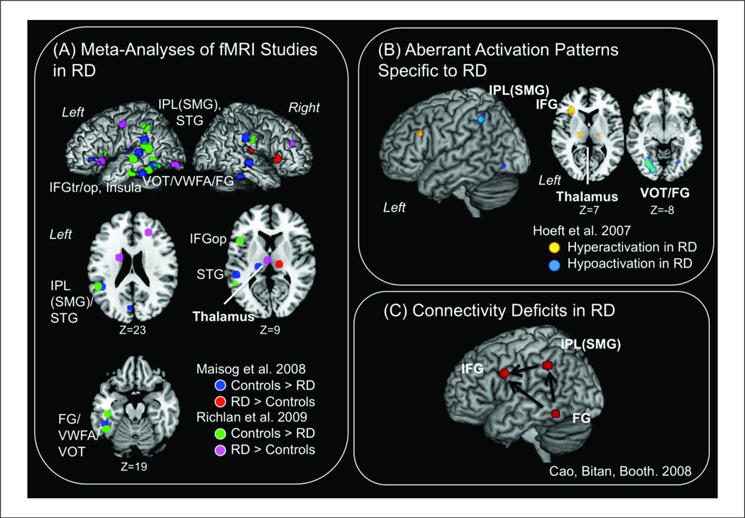
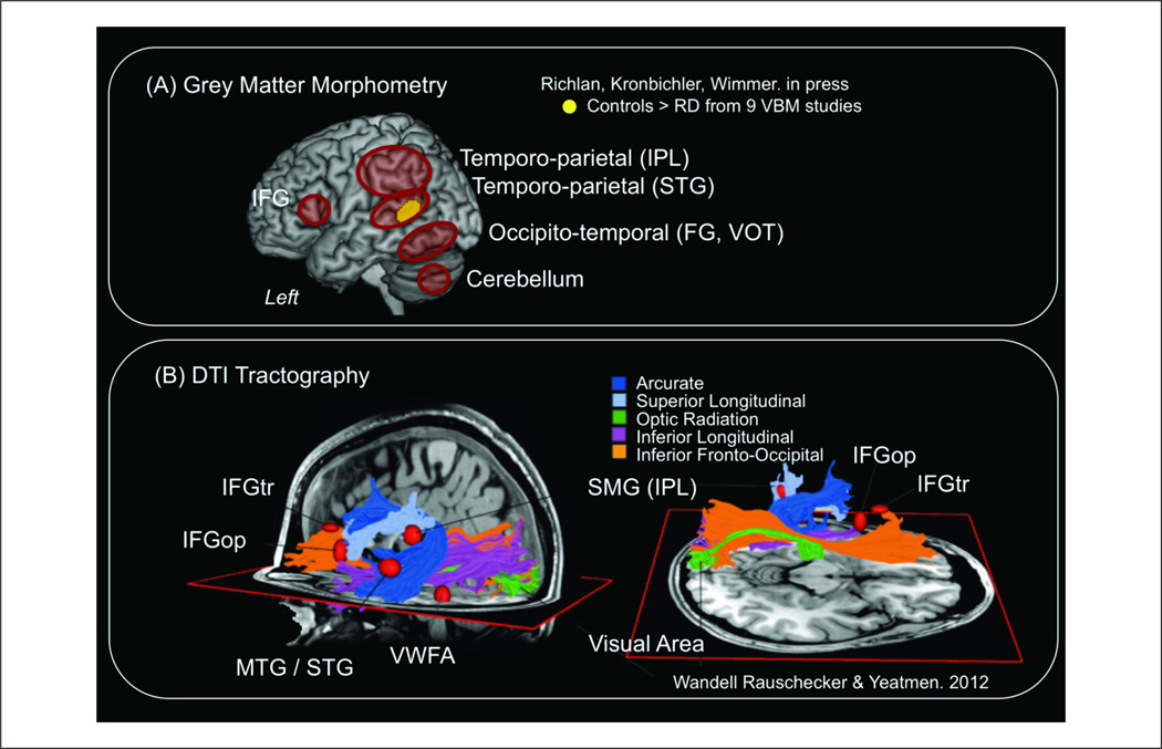
The primary goal of this review is to highlight current research and theories describing the neurobiological basis of math (MD), reading (RD), and comorbid math and reading disability (MD+RD). We first describe the unique brain and cognitive processes involved in acquisition of math and reading skills, emphasizing similarities and differences in each domain. Next we review functional imaging studies of MD and RD in children, integrating relevant theories from experimental psychology and cognitive neuroscience to characterize the functional neuroanatomy of cognitive dysfunction in MD and RD. We then review recent research on the anatomical correlates of MD and RD. Converging evidence from morphometry and tractography studies are presented to highlight distinct patterns of white matter pathways which are disrupted in MD and RD. Finally, we examine how the intersection of MD and RD provides a unique opportunity to clarify the unique and shared brain systems which adversely impact learning and skill acquisition in MD and RD, and point out important areas for future work on comorbid learning disabilities.

Keywords: comorbidity; learning disabilities; neurobiological.


Conflict of interest statement

Declaration of Conflicting Interests

The author(s) declared no potential conflicts of interest with respect to the research, authorship, and/or publication of this article.

Figures

Figure 1
Figure 1
Neurobiological pathways underlying math and reading disability (MD+RD) comorbidity. Schematic illustration of putative neurobiological pathways underlying MD+RD. (A) The “additive” pathway for comorbidity posits that pure RD and MD originate from unique cognitive and brain impairments, and the comorbid (“combined”) condition is the additive effect of impairment to brain systems that characterize MD and RD. (B) The “verbally mediated” pathway for comorbidity arises from the close link between symbolic mapping, fact retrieval, and word-based problem solving on one hand and verbally mediated processes on the other. In this case, comorbidity originates in impairments to phonological systems associated with RD. (C) The “domain-general” pathway for comorbidity originates from attention, working memory, and sequencing impairments such as those mediated by the prefrontal cortex (VLPFC and DLPFC). Again, this model states that pure RD and MD originate from unique cognitive and brain impairments. DLPFC = dorsolateral PFC; FG = fusiform gyrus; IPS = intraparietal sulcus; MTG = middle temporal gyrus; PFC = prefrontal cortex; SPL = superior parietal lobule; VLPFC = ventrolateral PFC.
Figure 2
Figure 2
Core math-related functional circuits. The intra-parietal sulcus (IPS) is a critical processing region in the brain for math and magnitude-related functions, and fronto-parietal circuits have been identified using resting-state functional connectivity analysis of the IPS. (A) IPS region-of -interests (ROIs) derived from cytoarchitectonic maps for the three sub-divisions of the IPS: hIP2 is the lateral and anterior sub-division of the IPS (blue); hIP1 is the sub-division located posterior to hIP2 (green); hIP3 is the posterior sub-division of the IPS (red). (B) Functional connectivity maps associated with hIP1, hIP2, and hIP3. The color code represents voxels correlated with each source ROI. The IPS has significant connectivity with distributed frontal (MFG and PMC) and parietal (SPL, and SMG) cortical regions in both hemispheres. Additional functional circuits associated with the ventral-occipital temporal cortex are not shown, but see Figure 3. Adapted from Uddin et al., 2010. hIP: human intra parietal; MFG: Middle frontal gyrus, PMC: PreMotor cortex, SMG: Supramarginal gyrus, SPL: Superior parietal lobule.
Figure 3
Figure 3
Aberrant functional brain responses during numercial tasks in MD. Brain regions that show decreased activation in MD relative to TD during (A) non-symbolic comparsion (Price et al. 2007), (B) symbolic comparison (Mussolin et al. 2010), and (C) an arithmetic problem solving task (Ashkenazi et al. 2012). Task paradigms are presented on the left in panels A, B, and C, with numerically “easy” tasks on top and numerically “difficult” tasks on the bottom. All studies in panels A through C found reduced activity in children with MD related to the difficulty of the tasks. (D) Summary of anatomical locations of brain regions highlighted in panels A through C. FG: Fusiform gyrus, IFG: Inferior frontal gyrus, IPS: Intraparietal sulcus, LOC: lateral occipital cortex; MFG: Middle frontal gyrus, MTG: Middle temporal gyrus, SPL: Superior parietal lobule.
Figure 4
Figure 4
Neuroanatomical substrates of MD. Neuroanatomical studies have shown that children with MD have reduced (A) grey matter volume in bilateral SPL, right IPS, lingual and fusiform gyri, MTL, and hippocampus, (B) white matter volume in right temporal-parietal cortex, and (C) lower fiber density in inferior longitudinal fasciculus, inferior fronto-occipital fasciculus, and caudal forceps major compared to typically developing children. The right panel of (C) shows reduced connectivity in children with MD for long-range white matter projection fibers that link the right fusiform gyrus with temporal-parietal areas (Adapted from Rykhlevskaia et al. 2009). IPS: Intraparietal sulcus, MTL: Middle temporal lobule, SPL: Superior parietal lobule. VBM: Voxel-based morphometry
Figure 5
Figure 5
Core reading-related functional circuits. (A) Surface rendering of 730 activation peaks reported in two meta-analyses of reading (Koyama et al., 2011; Vigneau et al., 2006). Each activation peak is color-coded according to its contrast category: phonology (blue), semantic (red), and syntax (green). Results of meta-analyses show a concentration of peaks in ventral occipito-temporal cortex (VOT), temporo-parietal region, inferior frontal cortex, and premotor/motor regions with overlapping representation for phonology, semantic and syntax processing. (B) Major reading-related functional circuit identified using resting-state functional connectivity with a seed (region of interest, ROI) placed in the fusiform gyrus (FG) within the VOT and Visual Word Form Area (VWFA). Connectivity from FG to the left inferior parietal lobule (IPL)/supramarginal gyrus (SMG) and pars opercularis (IFGop) of the left inferior frontal gyrus showed positive association with reading measures in typical adult readers (MNI coordinates derived from Koyama et al., 2011).
Figure 6
Figure 6
Aberrant functional brain activation during reading tasks in RD. (A) Brain regions that show under- and over-activation during fMRI tasks in RD adults and children compared to controls are color-coded (MNI coordinates taken from (Maisog, Einbinder, Flowers, Turkeltaub, & Eden, 2008; Richlan, Kronbichler, & Wimmer, 2009)). (B) Cold colors indicate brain regions that show reduced activation during a phonological processing fMRI task in RD compared to chronological age-matched and reading level-matched typical readers (i.e., RD-specific regions). Warm colors indicate brain regions that show increased activation in RD compared to age-matched but not reading-matched controls, likely reflecting compensatory mechanisms (Hoeft et al., 2007). (C) Connectivity analysis using direct causal modeling showed left FG to IPL and left FG/IPL to IFG connectivity deficits in RD (Cao, Bitan, & Booth, 2008). FG: Fusiform gyrus, IFGop: Inferior frontal gyrus, pars opercularis, IFGtr: IFG pars triangularis, IPL: Inferior parietal lobule, SMG: Supramarginal gyrus, STG: Superior temporal gyrus, VOT: Ventral occipito-temporal cortex, VWFA: Visual word form area.
Figure 7
Figure 7
Neuroanatomical substrates of RD. (A) Neuroanatomical studies have shown that children with RD have reduced grey matter (volume, density, and surface area) in left IFG, temporo-parietal region including IPL and superior temporal gyrus (STG), ventral occipitotemporal region (VOT) and cerebellum. Clusters in bilateral temporo-parietal region/ STG shown in orange have consistently reduced grey matter in RD compared to controls as reported in a recent meta-analysis (Richlan et al., in press). (B) White matter pathways that carry essential reading signals. The two images show several major white matter fascicles in the left hemisphere from different points of view. Three of these fascicles communicate information to and from the occipital lobe. The red ellipsoids are located in cortical regions activated in fMRI tasks of reading (Jobard, Crivello, & Tzourio-Mazoyer, 2003) (Reproduced with permission from (Wandell, Rauschecker, & Yeatman, 2012)). These pathways have been reported to be aberrant in RD in previous neuroimaging studies (except for the optic radiation) (Frye et al., 2011). IFGop: Inferior frontal gyrus, pars opercularis, IFGtr: IFG, pars triangularis, IPL: Inferior parietal lobule, MTG / STG: Middle temporal gyrus / superior temporal gyrus, SMG: Supramarginal gyrus, VWFA: Visual word form area.

References

    1. Abrams DA, Nicol T, Zecker SG, Kraus N. Auditory brainstem timing predicts cerebral asymmetry for speech. Journal of Neuroscience. 2006;26(43):11131–11137. - PMC - PubMed
    1. Abrams DA, Nicol T, Zecker S, Kraus N. Abnormal cortical processing of the syllable rate of speech in poor readers. Journal of Neuroscience. 2009;29(24):7686–7693. - PMC - PubMed
    1. Ahissar M. Dyslexia and the anchoring-deficit hypothesis. Trend in Cognitive Science. 2007;11(11):458–465. - PubMed
    1. Ansari D. Effects of development and enculturation on number representation in the brain. Nature Review Neuroscience. 2008;9(4):278–291. - PubMed
    1. Ansari D, Dhital B. Age-related changes in the activation of the intraparietal sulcus during nonsymbolic magnitude processing: An event-related functional magnetic resonance imaging study. Journal of Cognitive Neuroscience. 2006;18(11):1820–1828. - PubMed

Publication types