. 2013 May 15;32(10):1469-77.
doi: 10.1038/emboj.2013.82. Epub 2013 Apr 9.

Structure of the Legionella effector AnkX reveals the mechanism of phosphocholine transfer by the FIC domain

Affiliations

Structure of the Legionella effector AnkX reveals the mechanism of phosphocholine transfer by the FIC domain

Valérie Campanacci et al. EMBO J. .

Abstract

The FIC motif and the eukaryotic-like ankyrin repeats are found in many bacterial type IV effectors, yet little is known about how these domains enable bacteria to modulate host cell functions. Bacterial FIC domains typically bind ATP and transfer adenosine monophosphate moiety onto target proteins. The ankyrin repeat-containing protein AnkX encoded by the intracellular pathogen Legionella pneumophila is unique in that its FIC domain binds to CDP-choline and transfers a phosphocholine residue onto proteins in the Rab1 GTPase family. By determining the structures of unbound AnkX and AnkX with bound CDP-choline, CMP/phosphocholine and CMP, we demonstrate that the orientation of substrate binding in relation to the catalytic FIC motif enables this protein to function as a phosphocholinating enzyme rather than a nucleotidyl transferase. Additionally, the structure reveals that the ankyrin repeats mediate scaffolding interactions that resemble those found in protein-protein interactions, but are unprecedented in intramolecular interactions. Together with phosphocholination experiments, our structures unify a general phosphoryl transferase mechanism common to all FIC enzymes that should be conserved from bacteria to human.


Conflict of interest statement

The authors declare that they have no conflict of interest.

Figures

Figure 1
Figure 1
Structure of L. pneumophila AnkX. (A) Structure of AnkX with bound CDP-choline (in green). The sequences of the CMP binding, FIC, insert and ankyrin repeat domains are given in Supplementary Figure 1A. (B) Overlay of the four ankyrin repeats of AnkX1–484 (in magenta) to an archetypal ankyrin repeats protein (AnkyrinR, in orange Michaely et al, 2002). The longer loop in the first ankyrin motif is shown by an arrow. (C) The ankyrin repeats (in magenta) form extensive intramolecular contacts with the CMP-binding domain (in orange), the FIC domain (in cyan), and the insert domain (in red). Intramolecular contacts are shown in Supplementary Figure 3.
Figure 2
Figure 2
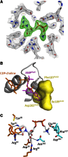
Structure of the CDP-choline binding site of AnkX. (A) Fo-Fc electron density omit map of CDP-choline bound to AnkXH229A contoured at 2.5σ. (B) Superposition of CDP-choline bound to AnkX (in orange) and AMP bound to cdc42 in the IbpA-cdc42 complex (in magenta, PDB 3N3V). Note that the AnkX active site (yellow surface) cannot accommodate the base of AMP. (C) Interactions of the CMP domain (in orange) and FIC domain (in cyan) residues with the cytidine and choline moieties of CDP-choline. Hydrogen bonds are shown in dotted lines.
Figure 3
Figure 3
Analysis of Rab1 phosphocholination and AnkX auto-phosphocholination. (A) AnkX constructs carrying mutations in the CDP-choline binding site have impaired Rab1 phosphocholination. Rab1 phosphocholination is expressed as the relative percentage of Rab1 phosphocholination by wild-type AnkX. Data are from immunoblots from in vitro reactions that contained full-length His-tagged AnkX constructs and Rab1A in the presence of phosphocholination buffer. Blots were probed with an anti-PC antibody. Representative blots are shown in Supplementary Figure 5A. Errors bars represent the standard deviation based on three independent experiments. (B) AnkX constructs carrying mutations in the FIC motif have impaired Rab1 phosphocholination. Experimental conditions are as in Figure 3A. Representative blots are shown in Supplementary Figure 5B. Errors bars represent the standard deviation based on three independent experiments. (C) Mg2+ is necessary for phosphocholination. Full-length His-tagged AnkX was dialysed against a metal-free buffer prior to the experiment, and subsequently assessed for auto-phosphocholination without (left lane) or with (right lane) addition of 1 mM Mg2+ in the auto-phosphocholination buffer. (D) Auto-phosphocholinated residues are not located in the FIC domain. The AnkX1–484 construct (lane 1) phosphocholinates Rab1 but is not auto-phosphocholinated. The AnkX1–484 construct carrying the H229A mutation is shown as a control that Rab1 phosphocholination requires the FIC motif histidine (lane 2). Molecular weight markers (in kDa) are shown on the right.
Figure 4
Figure 4
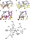
Structural basis for the phosphocholination reaction. (A) Interactions of the phosphates of CDP-choline with AnkXH229A. His229 in the FIC motif is overlaid from wild-type AnkX. (B) Interactions of the phosphates of CMP and phosphocholine with wild-type AnkX. (C) The Oαβ-Pβ bond of CDP-choline aligns with the phosphotyrosine bond of Cdc42 in complex with IbpA. (D) Proposed interactions of Mg2+ with CDP-choline modelled from Bartonella BepA/PPi-Mg2+ (PDB 2JK8). PPi bound to BepA is overlaid (in light blue). (E) Model for the transition state of phosphoryl transfer by the conserved FIC motif. Hydrogen bonds are in dotted lines, partial bonds are in dashed lines. Superpositions in Figure 4A–D are based on the FIC motif.
Figure 5
Figure 5
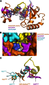
The active sites of FIC domains assemble from the conserved catalytic FIC motif and variable substrate-binding regions. (A) Superposition of active sites of phosphocholinating (AnkX, orange), AMPylating (IbpA, magenta) and unknown function (NmFic, blue and BepA, cyan) FIC enzymes. The FIC motif is shown in yellow and is in the same orientation in all views. Structural elements that recognize the leaving group are on the left, and the regions that recognize the transferred phosphoryl group are on the right. The latter regions are probably also involved in binding target proteins, as shown for the β-hairpin of IbpA (Xiao et al, 2010). (B) Overlaid surfaces of FIC enzymes active sites highlight the variety of their shapes and volumes. The FIC motif is shown in yellow. (C) Close-up view showing the common use of an aromatic residue by AnkX to bind the base ring of CDP-choline (in orange) and by the FIC-related AvrB protein (from PDB 2NUN) to bind ADP (in cyan). AMP bound to IbpA is overlaid (in magenta, PDB 3N3V).

Similar articles

Cited by

References

    1. Adams PD, Afonine PV, Bunkoczi G, Chen VB, Davis IW, Echols N, Headd JJ, Hung LW, Kapral GJ, Grosse-Kunstleve RW, McCoy AJ, Moriarty NW, Oeffner R, Read RJ, Richardson DC, Richardson JS, Terwilliger TC, Zwart PH (2010) PHENIX: a comprehensive Python-based system for macromolecular structure solution. Acta Crystallogr D Biol Crystallogr 66: 213–221 - PMC - PubMed
    1. Blanc E, Roversi P, Vonrhein C, Flensburg C, Lea SM, Bricogne G (2004) Refinement of severely incomplete structures with maximum likelihood in BUSTER-TNT. Acta Crystallogr D Biol Crystallogr 60: 2210–2221 - PubMed
    1. Cossart P, Roy CR (2010) Manipulation of host membrane machinery by bacterial pathogens. Curr Opin Cell Biol 22: 547–554 - PMC - PubMed
    1. Davis IW, Leaver-Fay A, Chen VB, Block JN, Kapral GJ, Wang X, Murray LW, Arendall WB 3rd, Snoeyink J, Richardson JS, Richardson DC (2007) MolProbity: all-atom contacts and structure validation for proteins and nucleic acids. Nucleic Acids Res 35: W375–W383 - PMC - PubMed
    1. Desveaux D, Singer AU, Wu AJ, McNulty BC, Musselwhite L, Nimchuk Z, Sondek J, Dangl JL (2007) Type III effector activation via nucleotide binding, phosphorylation, and host target interaction. PLoS Pathog 3: e48. - PMC - PubMed

Publication types