. 2013 May 31;288(22):16055-63.
doi: 10.1074/jbc.M113.456897. Epub 2013 Apr 9.

The structure of CcmP, a tandem bacterial microcompartment domain protein from the β-carboxysome, forms a subcompartment within a microcompartment

Affiliations

The structure of CcmP, a tandem bacterial microcompartment domain protein from the β-carboxysome, forms a subcompartment within a microcompartment

Fei Cai et al. J Biol Chem. .

Abstract

The carboxysome is a bacterial organelle found in all cyanobacteria; it encapsulates CO2 fixation enzymes within a protein shell. The most abundant carboxysome shell protein contains a single bacterial microcompartment (BMC) domain. We present in vivo evidence that a hypothetical protein (dubbed CcmP) encoded in all β-cyanobacterial genomes is part of the carboxysome. We show that CcmP is a tandem BMC domain protein, the first to be structurally characterized from a β-carboxysome. CcmP forms a dimer of tightly stacked trimers, resulting in a nanocompartment-containing shell protein that may weakly bind 3-phosphoglycerate, the product of CO2 fixation. The trimers have a large central pore through which metabolites presumably pass into the carboxysome. Conserved residues surrounding the pore have alternate side-chain conformations suggesting that it can be open or closed. Furthermore, CcmP and its orthologs in α-cyanobacterial genomes form a distinct clade of shell proteins. Members of this subgroup are also found in numerous heterotrophic BMC-associated gene clusters encoding functionally diverse bacterial organelles, suggesting that the potential to form a nanocompartment within a microcompartment shell is widespread. Given that carboxysomes and architecturally related bacterial organelles are the subject of intense interest for applications in synthetic biology/metabolic engineering, our results describe a new type of building block with which to functionalize BMC shells.

Keywords: CO2 Fixation; Carbon Dioxide; Carboxysome; Cell Compartmentation; Microbiology; Microcompartment; Protein Self-assembly; Protein Structure.


Figures

FIGURE 1.
FIGURE 1.
CcmP is a component of the β-carboxysome. A, organization of carboxysomal genes in S. elongatus PCC 7942 (Syn7942). Genes that contain BMC domain(s) are shown in red. B, wild-type cells harboring CcmP-cerulean and RbcL-GFP were visualized using laser scanning confocal microscopy following induction of Ptrc·CcmP-cerulean with 1 mm isopropyl 1-thio-β-d-galactopyranoside (IPTG) for 24 h. RbcL-GFP is constitutively expressed using the endogenous promoter upstream of ccmK2. The chlorophyll-a (chl-a) channel was used as a marker to identify the cell outline. Scale bar, 1 μm.
FIGURE 2.
FIGURE 2.
CcmP contains two BMC domains and forms a hexamer. A, CcmP hexamer shown in ribbons, colored by chain. B, monomer outlined in black was rotated 90° and rainbow-colored (blue, N terminus; red, C terminus) with 3PGA modeled in and shown as sticks. C, slab view of a CcmP hexamer, with one trimer (pseudo-hexamer) colored in green and one in cyan. The concave side of each trimer is indicated. D, when a map (σ of 1.3) was calculated without the presence of 3PGA molecule in the model, extra electron density was observed in the intra-domain pocket of a CcmP monomer near the conserved histidine residue (His-18). A 3PGA molecule (shown as sticks) can be credibly fit into this density.
FIGURE 3.
FIGURE 3.
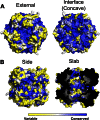
Sequence conservation mapped on the CcmP form 1 structure. Conservation scores were calculated with ConSurf (41). Each amino acid position is assigned a value from 1 (most variable) to 9 (most conserved) based on their evolutionary rates and then colored according to the grade with yellow variable and blue conserved. A, external side of pseudohexamer compared with interface (concave) side; B, side and slab view of hexamer, with 3PGA molecules shown as spheres and sticks.
FIGURE 4.
FIGURE 4.
CcmP packs in layers in the crystal. A, in the form 1 structure, hexamers of CcmP are packed in layers of alternating orientations of open (yellow) and closed (blue) trimers. The two side views of the layer show that strips with same orientations are on the same plane, although the alternately oriented strips are slightly shifted. B, transmission electron micrograph of CcmP. Some strips are indicated with red arrows. Scale bar size is 20 nm.
FIGURE 5.
FIGURE 5.
Phylogram of BMC domains. BMC domain containing proteins from α-carboxysomes (Cso, labels in black), β-carboxysomes (Ccm, labels in dark gray), and heterotrophic bacteria, where orthologs to CcmP are found (other, labels in light gray), are shown in an un-rooted distance tree. Two BMC domains (N-BMC and C-BMC) of CcmP, CsoS1D, and their heterotrophic orthologs are constructed separately. Bootstrap values were obtained from 100 replicates. Numbers above the branches indicate bootstrap values, and only branches receiving bootstrap values greater than 49 are indicated.

References

    1. Tanaka S., Kerfeld C. A., Sawaya M. R., Cai F., Heinhorst S., Cannon G. C., Yeates T. O. (2008) Atomic-level models of the bacterial carboxysome shell. Science 319, 1083–1086 - PubMed
    1. Kerfeld C. A., Sawaya M. R., Tanaka S., Nguyen C. V., Phillips M., Beeby M., Yeates T. O. (2005) Protein structures forming the shell of primitive bacterial organelles. Science 309, 936–938 - PubMed
    1. Kinney J. N., Axen S. D., Kerfeld C. A. (2011) Comparative analysis of carboxysome shell proteins. Photosynth. Res. 109, 21–32 - PMC - PubMed
    1. Tsai Y., Sawaya M. R., Yeates T. O. (2009) Analysis of lattice-translocation disorder in the layered hexagonal structure of carboxysome shell protein CsoS1C. Acta Crystallogr. D Biol. Crystallogr. 65, 980–988 - PubMed
    1. Tsai Y., Sawaya M. R., Cannon G. C., Cai F., Williams E. B., Heinhorst S., Kerfeld C. A., Yeates T. O. (2007) Structural analysis of CsoS1A and the protein shell of the Halothiobacillus neapolitanus carboxysome. PLoS Biol. 5, e144. - PMC - PubMed

Publication types

MeSH terms

LinkOut - more resources