

HPV-18 E6 mutants reveal p53 modulation of viral DNA amplification in organotypic cultures
- PMID: 23572574
- PMCID: PMC3651465
- DOI: 10.1073/pnas.1304855110
HPV-18 E6 mutants reveal p53 modulation of viral DNA amplification in organotypic cultures
Abstract
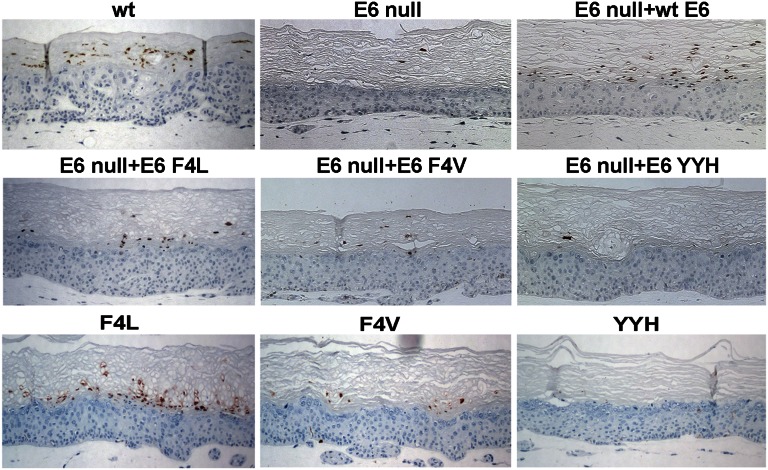
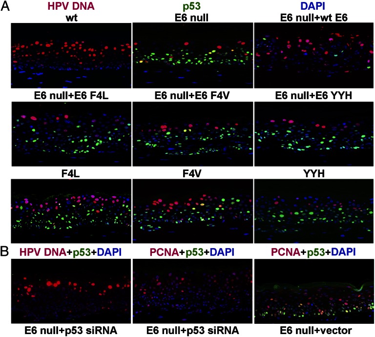
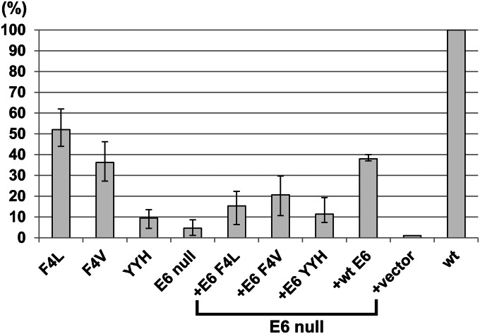
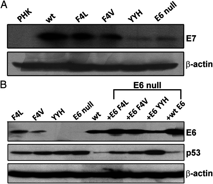
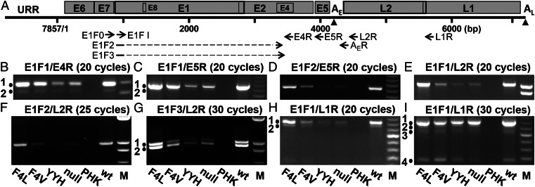
Human papillomaviruses (HPVs) amplify in differentiated strata of a squamous epithelium. The HPV E7 protein destabilizes the p130/retinoblastoma susceptibility protein family of tumor suppressors and reactivates S-phase reentry, thereby facilitating viral DNA amplification. The high-risk HPV E6 protein destabilizes the p53 tumor suppressor and many other host proteins. However, the critical E6 targets relevant to viral DNA amplification have not been identified, because functionally significant E6 mutants are not stably maintained in transfected cells. Using Cre-loxP recombination, which efficiently generates HPV genomic plasmids in transfected primary human keratinocytes, we have recapitulated a highly productive infection of HPV-18 in organotypic epithelial cultures. By using this system, we now report the characterization of four HPV-18 E6 mutations. An E6 null mutant accumulated high levels of p53 and amplified very poorly. p53 siRNA or ectopic WT E6 partially restored amplification, whereas three missense E6 mutations that did not effectively destabilize p53 complemented the null mutant poorly. Unexpectedly, in cis, two of the missense mutants amplified, albeit to a lower extent than the WT and only in cells with undetectable p53. These observations and others implicate p53 and additional host proteins in regulating viral DNA amplification and also suggest an inhibitory effect of E6 overexpression. We show that high levels of viral DNA amplification are critical for late protein expression and report several previously undescribed viral RNAs, including bicistronic transcripts predicted to encode E5 and L2 or an alternative form of E1^E4 and L1.
Keywords: HPV transcripts; human papillomavirus DNA amplification; trans complementation.
Conflict of interest statement
The authors declare no conflict of interest.
Figures
Similar articles
-
Robust production and passaging of infectious HPV in squamous epithelium of primary human keratinocytes.Genes Dev. 2009 Jan 15;23(2):181-94. doi: 10.1101/gad.1735109. Epub 2009 Jan 8. Genes Dev. 2009. PMID: 19131434 Free PMC article.
-
Vorinostat, a pan-HDAC inhibitor, abrogates productive HPV-18 DNA amplification.Proc Natl Acad Sci U S A. 2018 Nov 20;115(47):E11138-E11147. doi: 10.1073/pnas.1801156115. Epub 2018 Nov 1. Proc Natl Acad Sci U S A. 2018. PMID: 30385631 Free PMC article.
-
Human papillomavirus 16 E6 expression disrupts the p53-mediated cellular response to DNA damage.Proc Natl Acad Sci U S A. 1993 May 1;90(9):3988-92. doi: 10.1073/pnas.90.9.3988. Proc Natl Acad Sci U S A. 1993. PMID: 8387205 Free PMC article.
-
Cellular targets of the oncoproteins encoded by the cancer associated human papillomaviruses.Princess Takamatsu Symp. 1991;22:239-48. Princess Takamatsu Symp. 1991. PMID: 1668886 Review.
-
Interactions of HPV E6 and E7 oncoproteins with tumour suppressor gene products.Cancer Surv. 1992;12:197-217. Cancer Surv. 1992. PMID: 1322242 Review.
Cited by
-
Genome-Wide Transcriptome Analysis of Human Papillomavirus 16-Infected Primary Keratinocytes Reveals Subtle Perturbations Mostly due to E7 Protein Expression.J Virol. 2020 Jan 17;94(3):e01360-19. doi: 10.1128/JVI.01360-19. Print 2020 Jan 17. J Virol. 2020. PMID: 31748387 Free PMC article.
-
YAP1 activation by human papillomavirus E7 promotes basal cell identity in squamous epithelia.Elife. 2022 Feb 16;11:e75466. doi: 10.7554/eLife.75466. Elife. 2022. PMID: 35170430 Free PMC article.
-
Serine/Arginine-Rich Splicing Factor 3 and Heterogeneous Nuclear Ribonucleoprotein A1 Regulate Alternative RNA Splicing and Gene Expression of Human Papillomavirus 18 through Two Functionally Distinguishable cis Elements.J Virol. 2016 Sep 29;90(20):9138-52. doi: 10.1128/JVI.00965-16. Print 2016 Oct 15. J Virol. 2016. PMID: 27489271 Free PMC article.
-
The Role of the DNA Damage Response throughout the Papillomavirus Life Cycle.Viruses. 2015 May 21;7(5):2450-69. doi: 10.3390/v7052450. Viruses. 2015. PMID: 26008695 Free PMC article. Review.
-
Establishment of a Three-Dimensional In Vitro Model of Equine Papillomavirus Type 2 Infection.Viruses. 2021 Jul 19;13(7):1404. doi: 10.3390/v13071404. Viruses. 2021. PMID: 34372610 Free PMC article.
References
-
- de Villiers E-M, Fauquet C, Broker TR, Bernard H-U, zur Hausen H. Classification of papillomaviruses. Virology. 2004;324(1):17–27. - PubMed
-
- zur Hausen H. Papillomaviruses in the causation of human cancers—a brief historical account. Virology. 2009;384(2):260–265. - PubMed
-
- Chow LT, Broker TR, Steinberg BM. The natural history of human papillomavirus infections of the mucosal epithelia. APMIS. 2010;118(6–7):422–449. - PubMed
Publication types
MeSH terms
Substances
Grants and funding
LinkOut - more resources
Full Text Sources
Other Literature Sources
Molecular Biology Databases
Research Materials
Miscellaneous
