. 2013 Apr 30;110(18):E1676-84.
doi: 10.1073/pnas.1217712110. Epub 2013 Apr 9.

Transposon mutagenesis identifies genes essential for Plasmodium falciparum gametocytogenesis

Affiliations

Transposon mutagenesis identifies genes essential for Plasmodium falciparum gametocytogenesis

Hiromi Ikadai et al. Proc Natl Acad Sci U S A. .

Abstract

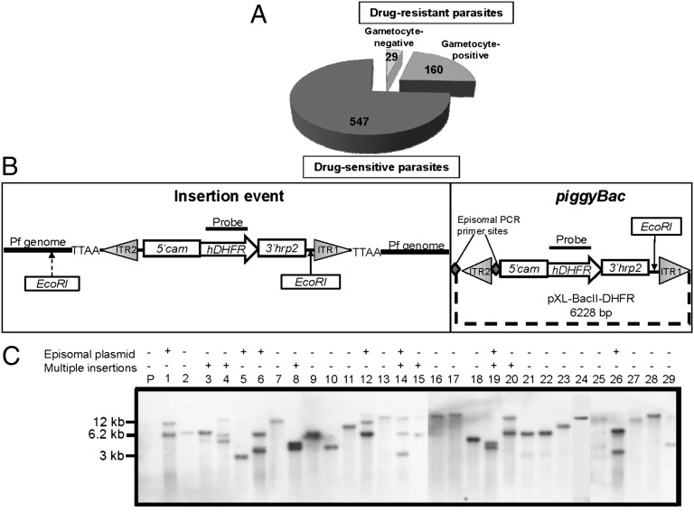
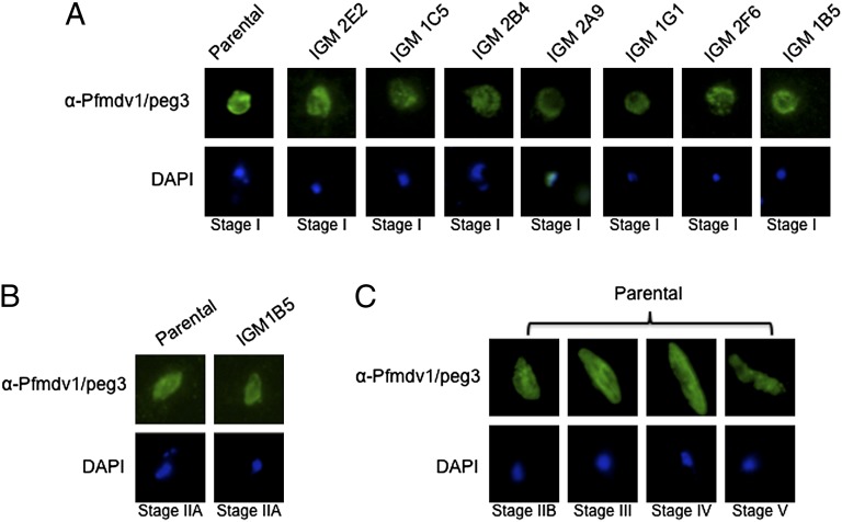
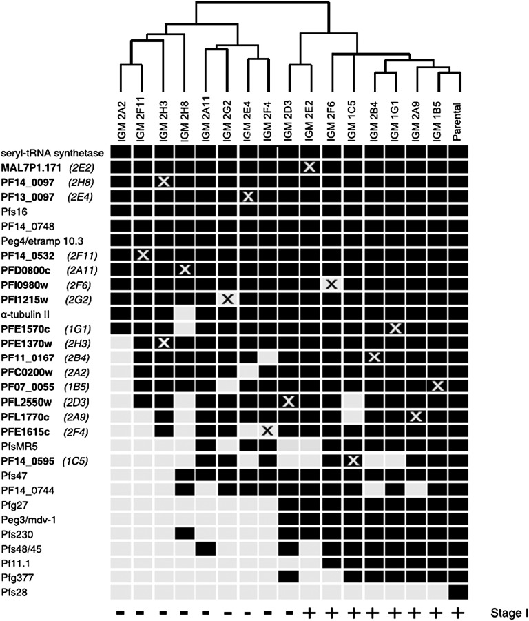
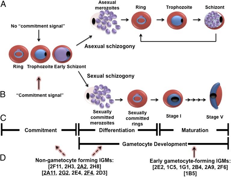
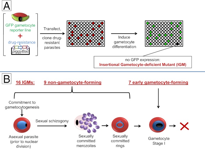
Gametocytes are essential for Plasmodium transmission, but little is known about the mechanisms that lead to their formation. Using piggyBac transposon-mediated insertional mutagenesis, we screened for parasites that no longer form mature gametocytes, which led to the isolation of 29 clones (insertional gametocyte-deficient mutants) that fail to form mature gametocytes. Additional analysis revealed 16 genes putatively responsible for the loss of gametocytogenesis, none of which has been previously implicated in gametocytogenesis. Transcriptional profiling and detection of an early stage gametocyte antigen determined that a subset of these mutants arrests development at stage I or in early stage II gametocytes, likely representing genes involved in gametocyte maturation. The remaining mutants seem to arrest before formation of stage I gametocytes and may represent genes involved in commitment to the gametocyte lineage.


Conflict of interest statement

The authors declare no conflict of interest.

Figures

Fig. 1.
Fig. 1.
piggyBac insertional mutagenesis results in 22 IGMs with single insertion events. (A) In three independent transfection experiments, 736 wells were seeded with parasites. Of these wells, 189 contained drug-resistant parasites. Of these wells, 29 (15%) failed to form gametocytes and were named IGMs. (B) Schematic diagram of the piggyBac plasmid and an insertion event. (Left) When digested with EcoRI and hybridized with an hdhfr probe, a Southern blot will yield a band with size that depends on the position of the closest EcoRI site in the P. falciparum genome. (Right) A nonintegrated episomal plasmid yields a 6.2-kb fragment (it has only one EcoRI site). ITR, transposon inverted terminal repeat. pXL-BacII-DHFR diagram was modified from ref. . PCR primer sites for the piggyBac plasmid are indicated by large gray diamonds. (C) Of 29 IGMs, 22 IGMs yielded a single novel band (different from the 6.2-kb band), indicating a single piggyBac insertion (lanes 2, 7, 9, 10, 11, 13, 16, 17, 18, 21, 22, 23, 24, 25, 27, 28, and 29). Some IGMs with integrated transposons also yielded an ∼6.2-kb band, suggesting that a nonintegrated episomal plasmid is still present in these parasites. PCR analysis was used to determine whether an episome is present in the mutant clone (Table S1) or a separate piggyBac integration event occurred in another region of the genome. IGMs with multiple insertion events (lanes 3, 4, 8, 14, 15, 19, and 20) were not further characterized. No signal was detected with DNA from parental parasites as expected.
Fig. 2.
Fig. 2.
Episomal complementation restores gametocytogenesis to five of the IGMs. (A) The rescue construct contains the endogenous putatively disrupted gene along with ∼1.5–2 kb upstream of the ATG translational initiator (5′) and ∼0.3–0.8 kb downstream (3′) of the stop codon. The construct confers resistance to G418/Geneticin. (B) As a control, the IGMs and the parental line were also transfected with an empty vector, which only contains the Geneticin/G418 selectable marker. (C) RT-PCR showing that the complementation constructs restored mRNA expression to levels similar to parental parasites. CTRL, control seryl-tRNA synthetase; GSP, gene-specific primer. The bottom line shows that microscopic analysis revealed that complemented parasites produced gametocytes in numbers comparable with the parental line, whereas IGMs transfected with the empty construct did not produce gametocytes. Gametocyte numbers were assayed three independent times, and each time, 10,000 RBCs were counted. Except for IGM 2D3, differences in gametocyte numbers between the rescued IGMs and the parental line were not statistically significant (Poisson). Whereas IGM 2D3 transfected with the rescue vector produced target mRNA of abundance comparable to the parental line, it did not produce gametocytes, suggesting that a second site mutation accounts for impaired gametocytogenesis.
Fig. 3.
Fig. 3.
A subset of the IGMs forms stage I gametocytes. (A) Pfmdv1/peg3 is a gametocyte-specific gene expressed in all gametocyte stages (13). Immunofluorescence using antisera against Pfmdv1/peg3 identified the protein in stage I gametocytes from IGMs 2E2, 1C5, 2B4, 1G1, 2F6, and 1B5. (B) Pfmdv1/peg3 staining also reveals that IGM 1B5 is able to produce what appears to be stage IIA gametocytes but not stage IIB or later gametocytes (8). None of the IGMs produced gametocytes that mature beyond stage I or IIA. (C) Under the same experimental conditions, the antibody consistently identified the protein in stages I–V gametocytes from the parental line.
Fig. 4.
Fig. 4.
Hierarchical ordering of the IGMs by RT-PCR expression profiling. The expression profiles indicate transcripts that were expressed (black boxes) or not expressed (light gray boxes) as measured by semiquantitative RT-PCR (Fig. S3). The transcripts listed correspond to 16 putatively disrupted genes as well as known early (Pfs16, PF14_0748, Peg4/etramp10.3, Pfg27, PF14_0744, and Pfmdv1/peg3) and late (Pf11.1, Pfg377, Pfs48/45, PfMR5, Pfs47, and Pfs28) gametocyte-specific genes. An X indicates the transcript from the putatively disrupted IGM gene. Transcript abundance from some putatively disrupted genes was only decreased and not completely absent in some IGMs (gray Xs on black boxes). The IGMs are hierarchically clustered based on the similarity of their expression profiles to each other and the parental line. All IGMs that are able to differentiate into stage I gametocytes and express the Pfmdv1/peg3 marker (Fig. 3, indicated along the bottom) cluster to the right next to the WT parental line. Gametocyte-specific transcript accession numbers are in Table S3.
Fig. 5.
Fig. 5.
Model for gametocyte commitment, differentiation, and maturation. P. falciparum has two developmental fates: (A) cyclic asexual propagation or (B) terminal sexual differentiation. (A) In humans, the majority of the blood stage parasites undergo asexual propagation (default pathway). Here, the parasite undergoes asexual schizogny, producing asexual merozoites that, when released, invade new erythrocytes and continue the cycle every 48 h. The asexual propagation of the parasite accounts for disease symptoms such as cyclic fever. (B) The parasite undergoes sexual differentiation (gametocytogenesis) if, before DNA replication, a commitment signal is received (the position of the up arrow is arbitrary, because the timing of commitment is unknown). This commitment signal is ill defined, but in vitro, it is typically associated with environmental conditions that negatively impact asexual reproduction, such as high parasitemia or changes in hematocrit (2, 31). After committed, the parasite initiates gametocyte development through sexual schizogony that results in formation of sexually committed merozoites. When released, the committed merozoites invade fresh erythrocytes to form sexually committed rings. There are currently no known markers for this initial process. During the next ∼24 h, the parasite differentiates into a morphologically and molecularly distinguishable stage I gametocyte. Thereafter, the gametocyte undergoes a stepwise maturation process to yield a mature stage V male or female gametocyte. Only stage V gametocytes circulate in the bloodstream, and this stage is the only stage that is infectious to the Anopheles mosquito vector. (C) Gametocytogenesis can be divided into three steps: commitment, prestage I gametocyte development, and poststage I gametocyte development. Commitment is the process that commits the parasite to sexual differentiation. Prestage I gametocyte development begins with sexual schizogony and concludes when the committed ring stage parasite transforms into an identifiable stage I gametocyte (∼24–30 h postinvasion). Poststage I gametocyte development is the process that transforms a stage I gametocyte into a mature stage V gametocyte. (D) One group of IGMs does not form stage I gametocytes. These mutants likely arrest during commitment prestage I development. Because there are no known markers that discriminate between the two processes, at present, there are no means to determine at which point these IGMs arrest (as indicated by the dashed arrows). A second group of IGMs forms stage I gametocytes but then arrests (as indicated by the solid arrow). The genes disrupted in these IGMs likely play a role in gametocyte maturation. The underlined IGMs are the complemented lines.
Fig. P1.
Fig. P1.
(A) A P. falciparum reporter line was created that carries a GFP gene driven by a late stage gametocyte-specific promoter (Pfs28). Reporter parasites were transfected with a piggyBac transposon containing a drug-selectable marker. Drug-resistant clones were grown under conditions that favored gametocyte differentiation, and 16 rare clones that did not produce GFP-positive gametocytes (IGMs) were found to carry single transposon insertions at different chromosomal locations. (B) After committed, the parasite undergoes sexual schizogony, producing merozoites that rupture and invade fresh erythrocytes to form a sexually committed ring. At ∼24 h postinvasion, the parasite can be identified as a stage I gametocyte that develops into a mature stage V gametocyte. Seven IGMs formed stage I gametocytes and then arrested, likely because the disrupted loci in these IGMs are essential for maturation. The nine remaining IGMs did not form identifiable gametocytes, likely because the disrupted loci play an essential role in commitment or differentiation.

Similar articles

Cited by

References

    1. Bousema T, Drakeley C. Epidemiology and infectivity of Plasmodium falciparum and Plasmodium vivax gametocytes in relation to malaria control and elimination. Clin Microbiol Rev. 2011;24(2):377–410. - PMC - PubMed
    1. Carter R, Miller LH. Evidence for environmental modulation of gametocytogenesis in Plasmodium falciparum in continuous culture. Bull World Health Organ. 1979;57(Suppl 1):37–52. - PMC - PubMed
    1. Inselburg J. Gametocyte formation by the progeny of single Plasmodium falciparum schizonts. J Parasitol. 1983;69(3):584–591. - PubMed
    1. Bruce MC, Alano P, Duthie S, Carter R. Commitment of the malaria parasite Plasmodium falciparum to sexual and asexual development. Parasitology. 1990;100(Pt 2):191–200. - PubMed
    1. Silvestrini F, Alano P, Williams JL. Commitment to the production of male and female gametocytes in the human malaria parasite Plasmodium falciparum. Parasitology. 2000;121(Pt 5):465–471. - PubMed

Publication types

MeSH terms

Substances