Stereological study of the diabetic heart of male rats
- PMID: 23573103
- PMCID: PMC3616204
- DOI: 10.5625/lar.2013.29.1.12
Stereological study of the diabetic heart of male rats
Abstract
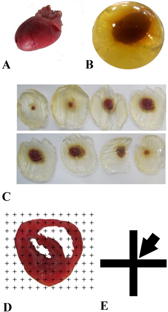
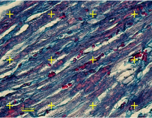
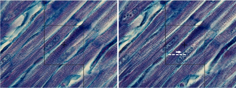
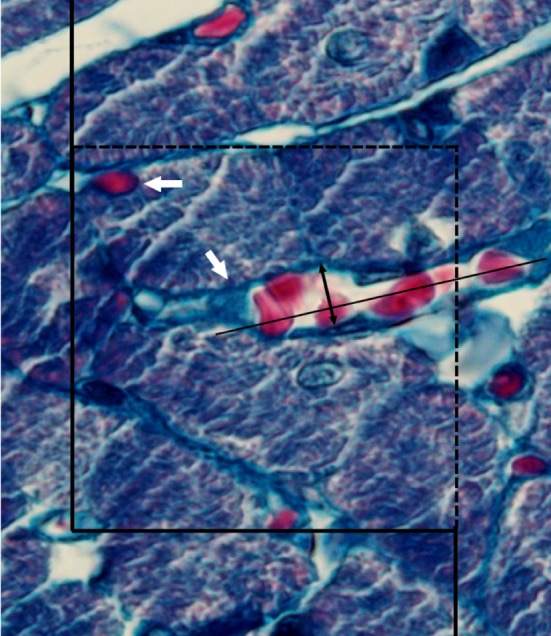
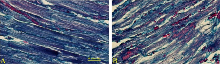
The present study aimed to quantitatively compare the normal and diabetic hearts of rats using stereological methods. Diabetic and control rats received streptozotocin (60 mg/kg) and no treatments, respectively. On the 56(th) day, the hearts were removed and their total volume was estimated using isotropic Cavalieri method. The total volume of the connective tissues and vessels, total length and diameter of the vessels, total number of cardiomyocytes nuclei, and the mean volume of the cardiomyocytes were estimated, as well. In comparison to the control animals, 60 and 43% increase was observed in the total volume of the connective tissue and microvessels of the diabetic rats, respectively (P<0.05). The percent of the vessel profiles with the diameter of 2-4 µm was decreased, while the percent of the vessel profiles with the diameter of 4.1-8 µm was increased in the diabetic hearts (P<0.05). No significant difference was found in the vessels with more than 8 µm diameters. The total number of the cardiomyocytes' nuclei and the number-weighted mean volume were respectively decreased by 37 and 64% in the diabetic group (P<0.01). A significant difference was observed between the two groups concerning the left ventricle volume to body weight ratio as an index for ventricular hypertrophy (P<0.05), while no difference was found regarding the right ventricle to body weight ratio. It can be concluded that diabetes can induce structural changes, including loss and/or atrophy of the cardiomyocytes, accompanied with increase in the connective tissue in the rats' hearts.
Keywords: Stereology; diabetes; heart.
Figures
Similar articles
-
Stereology of the myocardium and blood biochemistry in aged rats fed with a cholesterol-rich and canola oil diet (n-3 fatty acid rich).Basic Res Cardiol. 1998 Jun;93(3):182-91. doi: 10.1007/s003950050085. Basic Res Cardiol. 1998. PMID: 9689444
-
Spatial arrangement of the heart structure: Application of second-order stereology in diabetic rats.Ann Anat. 2014 Jan;196(1):20-5. doi: 10.1016/j.aanat.2013.04.012. Epub 2013 May 23. Ann Anat. 2014. PMID: 23773475
-
Microscopic evaluation of the ventricular tissue using stereological and Voronoi tessellation methods: Application on doxorubicin-induced cardiotoxicity in rats.Micron. 2017 Oct;101:1-7. doi: 10.1016/j.micron.2017.05.004. Epub 2017 May 24. Micron. 2017. PMID: 28558328
-
Prolonged ejection duration helps to maintain pump performance of the renal-hypertensive-diabetic rat heart: correlations between isolated papillary muscle function and ventricular performance in situ.Cardiovasc Res. 1997 Apr;34(1):230-40. doi: 10.1016/s0008-6363(96)00239-8. Cardiovasc Res. 1997. PMID: 9217895
-
Cardiac hypertrophy in diabetic spontaneously hypertensive rats: role of angiotensin II?Clin Exp Pharmacol Physiol. 1997 Jun;24(6):445-8. doi: 10.1111/j.1440-1681.1997.tb01221.x. Clin Exp Pharmacol Physiol. 1997. PMID: 9171955
Cited by
-
Protective effects of aerobic exercise on cardiac histology and stereological parameters in a rat model of type 2 diabetes mellitus.J Diabetes Metab Disord. 2025 Jun 2;24(1):138. doi: 10.1007/s40200-025-01641-5. eCollection 2025 Jun. J Diabetes Metab Disord. 2025. PMID: 40469910
References
-
- Cheng YZ, Chen LJ, Lee WJ, Chen MF, Jung Lin H, Cheng JT. Increase of myocardial performance by Rhodiola-ethanol extract in diabetic rats. J Ethnopharmacol. 2012;144(2):234–239. - PubMed
-
- Kannel WB, Hjortland M, Castelli WP. Role of diabetes in congestive heart failure: the Framingham study. Am J Cardiol. 1974;34(1):29–34. - PubMed
-
- Abbott RD, Donahue RP, Kannel WB, Wilson PW. The impact of diabetes on survival following myocardial infarction in men vs women. The Framingham Study. JAMA. 1988;260(23):3456–3460. - PubMed
-
- Rubler S, Dlugash J, Yuceoglu YZ, Kumral T, Branwood AW, Grishman A. New type of cardiomyopathy associated with diabetic glomerulosclerosis. Am J Cardiol. 1972;30(6):595–602. - PubMed
LinkOut - more resources
Full Text Sources
Other Literature Sources