. 2013:2013:363076.
doi: 10.1155/2013/363076. Epub 2013 Mar 14.

Evaluation of In Vitro Anti-Inflammatory Activities and Protective Effect of Fermented Preparations of Rhizoma Atractylodis Macrocephalae on Intestinal Barrier Function against Lipopolysaccharide Insult

Affiliations

Evaluation of In Vitro Anti-Inflammatory Activities and Protective Effect of Fermented Preparations of Rhizoma Atractylodis Macrocephalae on Intestinal Barrier Function against Lipopolysaccharide Insult

Shambhunath Bose et al. Evid Based Complement Alternat Med. 2013.

Abstract

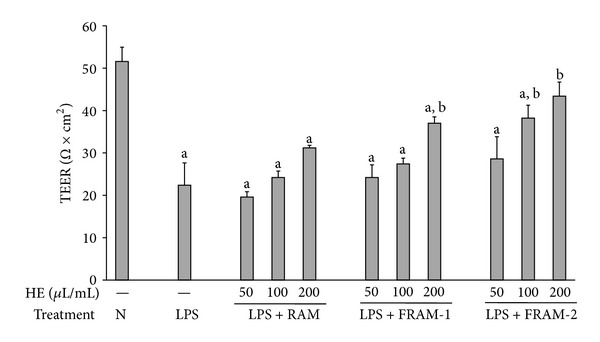
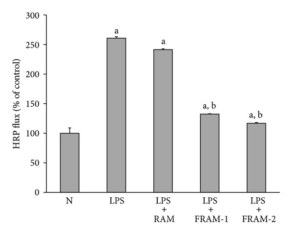
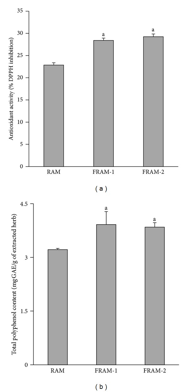
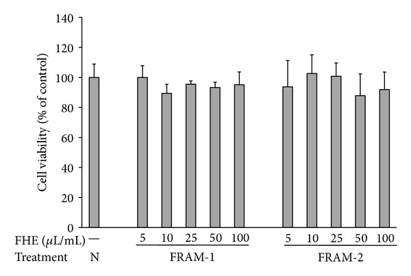
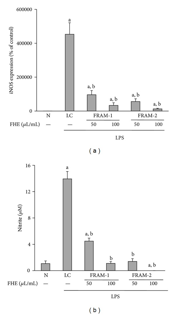
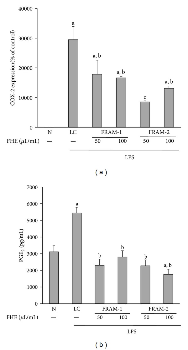
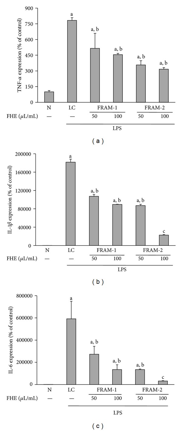
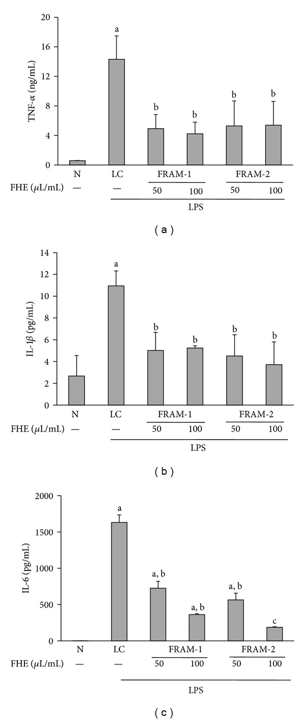
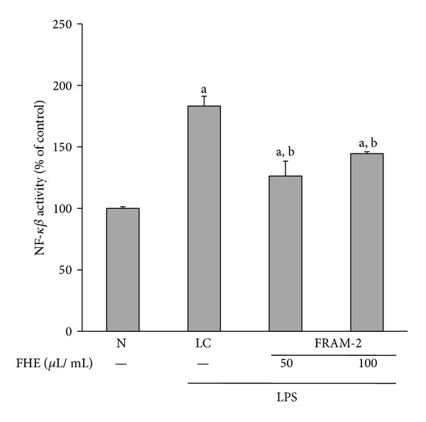
Lipopolysaccharide (LPS), a potent inducer of systemic inflammatory responses, is known to cause impairment of intestinal barrier function. Here, we evaluated the in vitro protective effect of an unfermented formulation of Rhizoma Atractylodis Macrocephalae (RAM), a traditional Chinese herbal medicine widely used in the treatment of many digestive and gastrointestinal disorders, and two fermented preparations of RAM, designated as FRAM-1 (prepared in Luria-Bertani broth) and FRAM-2 (prepared in glucose), on intestinal epithelial cells (IECs) against LPS insult. In general, fermented formulations, especially FRAM-2, but not unfermented RAM, exerted an appreciable protective effect on IECs against LPS-induced perturbation of membrane resistance and permeability. Both fermented formulations exhibited appreciable anti-inflammatory activities in terms of their ability to inhibit LPS-induced gene expression and induced production of a number of key inflammatory mediators and cytokines in RAW 264.7 macrophage cells. However, in most cases, FRAM-2 exhibited stronger anti-inflammatory effects than FRAM-1. Our findings also suggest that suppression of nuclear factor- κ β (NF- κ β ) activity might be one of the possible mechanisms by which the fermented RAM exerts its anti-inflammatory effects. Collectively, our results highlight the benefits of using fermented products of RAM to protect against LPS-induced inflammatory insult and impairment in intestinal barrier function.


Figures

Figure 1
Figure 1
The impact of treatment with saline or with different concentrations of unfermented Rhizoma Atractylodis Macrocephalae (RAM) or fermented RAM-1 (FRAM-1) and fermented RAM-2 (FRAM-2) formulations on the transepithelial electrical resistance (TEER) of HCT-116 cells exposed to LPS. The cells were treated with the indicated concentrations of RAM and FRAMs for 24 h. The control cells (N) and the noncontrol cells that were assigned to treatment with LPS alone (LPS) were treated with sterile saline instead of herbal extracts. After this treatment, the noncontrol and control cells were treated with LPS (10 μg/mL) and PBS, respectively, for 24 h, followed by the performance of TEER measurement. The detailed treatment regimen and experimental conditions are described in Section 2. The data are expressed as the mean ± SD, n = 3. aStatistically significant difference compared to control cells (P < 0.05); bstatistically significant difference compared to cells treated with LPS plus saline (P < 0.05). HE: herbal extract.
Figure 2
Figure 2
The impact of treatment with saline or with different concentrations of unfermented Rhizoma Atractylodis Macrocephalae (RAM) or fermented RAM-1 (FRAM-1) and fermented RAM-2 (FRAM-2) formulations on the HRP-flux of HCT-116 cells exposed to LPS. The cells were treated with RAM and FRAMs at a concentration of 200 μL/mL for 24 h. The control cells (N) and the noncontrol cells that were assigned to treatment with LPS alone were treated with sterile saline instead of herbal extracts. After this treatment, the noncontrol and control cells were treated with LPS (10 μg/mL) and PBS, respectively, for 24 h, followed by HRP-flux measurement. The detailed treatment regimen and experimental conditions are described in Section 2. The data are expressed as the mean ± SD, n = 3. The data were log-transformed prior to analysis by ANOVA. aStatistically significant difference compared to control cells (P < 0.05); bstatistically significant difference compared to cells treated with LPS plus saline (P < 0.05).
Figure 3
Figure 3
The DPPH radical scavenging activity (a) and total polyphenol content (b) of unfermented Rhizoma Atractylodis Macrocephalae (RAM) or fermented RAM-1 (FRAM-1) and fermented RAM-2 (FRAM-2) formulations. The detailed treatment regimen and experimental conditions are described in Section 2. The data are expressed as the mean ± SD, n = 3. aStatistically significant difference compared to RAM (P < 0.05).
Figure 4
Figure 4
The impact of treatment with saline (N) or with different concentrations of fermented Rhizoma Atractylodis Macrocephalae-1 (FRAM-1) or fermented Rhizoma Atractylodis Macrocephalae-2 (FRAM-2) formulations on the viability of RAW264.7 cells. The cells were treated for 24 h with the indicated concentrations of FRAMs followed by the assessment of cell viability. The detailed treatment regimen and experimental conditions are described in Section 2. The viability of the cells that were treated with saline was set to 100%. The data are expressed as the mean ± SD, n = 4. No statistically significant differences were observed between the treatment groups. FHE: fermented herbal extract.
Figure 5
Figure 5
The effect of treatment with LPS in combination with saline (LC) or with two different concentrations of fermented Rhizoma Atractylodis Macrocephalae-1 (FRAM-1) and fermented Rhizoma Atractylodis Macrocephalae-2 (FRAM-2) formulations on the expression of the iNOS gene (a) and production of nitrite (b) in RAW264.7 cells. The cells were treated for 24 h with the indicated concentrations of FRAMs. The control cells (N) and the noncontrol cells that were assigned to treatment with LPS alone (LC) were treated with sterile saline instead of herbal extracts. After this treatment, the noncontrol and control cells were treated with LPS (10 μg/mL) and PBS, respectively, for 24 h, followed by the determination of iNOS gene expression and nitrite production. The detailed treatment regimen and experimental conditions are described in Section 2. The level of iNOS gene expression in control cells (N) was set to 100%. The data are expressed as the mean ± SD, n = 3. The data for both iNOS and nitrite production were log-transformed prior to analysis by ANOVA. aStatistically significant difference compared to control cells (P < 0.05); bstatistically significant difference compared to cells treated with LPS plus saline (P < 0.05). FHE: fermented herbal extract.
Figure 6
Figure 6
The effect of treatment with LPS in combination with saline (LC) or with two different concentrations of fermented Rhizoma Atractylodis Macrocephalae-1 (FRAM-1) and fermented Rhizoma Atractylodis Macrocephalae-2 (FRAM-2) formulations on the expression of the COX-2 gene (a) and production of PGE2 (b) in RAW264.7 cells. The cells were treated with the indicated concentrations of FRAMs for 24 h. The control cells (N) and the noncontrol cells that were assigned to treatment with LPS alone (LC) were treated with sterile saline instead of herbal extracts. After this treatment, the noncontrol and control cells were treated with LPS (10 μg/mL) and PBS, respectively, for 24 h, followed by determination of COX-2 gene expression and PGE2 production. The detailed treatment regimen and experimental conditions are described in Section 2. The level of COX-2 gene expression in the control cells (N) was set to 100%. The data are expressed as the mean ± SD, n = 3. The data for COX-2 expression were log-transformed prior to analysis by ANOVA. aStatistically significant difference compared to control cells (P < 0.05); bstatistically significant difference compared to cells treated with LPS plus saline (P < 0.05); csignificantly lower compared to all other treatment groups (P < 0.05). FHE: fermented herbal extract.
Figure 7
Figure 7
The effect of treatment with LPS in combination with saline (LC) or with two different concentrations of fermented Rhizoma Atractylodis Macrocephalae-1 (FRAM-1) and fermented Rhizoma Atractylodis Macrocephalae-2 (FRAM-2) formulations on expression of TNF-α (a), IL-1β (b), and IL-6 (c) genes in RAW264.7 cells. The cells were treated with the indicated concentrations of FRAMs for 24 h. The control cells (N) and the noncontrol cells that were assigned to treatment with LPS alone (LC) were treated with sterile saline instead of herbal extracts. After this treatment, the noncontrol and control cells were treated with LPS (10 μg/mL) and PBS, respectively, for 24 h, followed by determination of gene expression of the above-mentioned cytokines. The detailed treatment regimen and experimental conditions are described in Section 2. The values are expressed as the mean ± SD, n = 3. The data for expression of all genes were log-transformed prior to analysis by ANOVA. aStatistically significant difference compared to control cells (P < 0.05); bstatistically significant difference compared to cells treated with LPS plus saline (P < 0.05); csignificantly lower compared to all other treatment groups (P < 0.05). FHE: fermented herbal extract.
Figure 8
Figure 8
The effect of treatment with LPS in combination with saline (LC) or with two different concentrations of fermented Rhizoma Atractylodis Macrocephalae-1 (FRAM-1) and fermented Rhizoma Atractylodis Macrocephalae-2 (FRAM-2) formulations on production of TNF-α (a), IL-1β (b), and IL-6 (c) by RAW264.7 cells. The cells were treated for 24 h with the indicated concentrations of FRAMs. The control cells (N) and the noncontrol cells that were assigned to treatment with LPS alone (LC) were treated with sterile saline instead of herbal extracts. After this treatment, the noncontrol and control cells were treated with LPS (10 μg/mL) and PBS, respectively, for 24 h; production of the above-mentioned cytokines was then determined. The detailed treatment regimen and experimental conditions are described in Section 2. The values are expressed as the mean ± SD, n = 3. The data for expression of the IL-6 gene were log-transformed prior to analysis by ANOVA. aStatistically significant difference compared to control cells (P < 0.05); bStatisticaly significant difference compared to cells treated with LPS plus saline (P < 0.05); csignificantly lower compared to all other treatment groups (P < 0.05). FHE, Fermented herbal extract.
Figure 9
Figure 9
The effect of treatment with LPS in combination with saline (LC) or with two different concentrations of fermented Rhizoma Atractylodis Macrocephalae-2 (FRAM-2) on NF-κβ activity in RAW264.7 cells. The cells were treated with the indicated concentrations of FRAM-2 for 24 h. The control cells (N) and the noncontrol cells that were assigned to treatment with LPS alone (LC) were treated with sterile saline instead of herbal extract. After this treatment, the noncontrol and control cells were treated with LPS (10 μg/mL) and PBS, respectively, for 1 h; the activation of NF-κβ was then determined. The detailed treatment regimen and experimental conditions are described in Section 2. The level of NF-κβ activity in the control cells (N) was set to 100%. The values are expressed as the mean ± SD, n = 3. The data were log-transformed prior to analysis by ANOVA. aStatistically significant difference compared to control cells (P < 0.05); bstatistically significant difference compared to cells treated with LPS plus saline (P < 0.05). FHE: fermented herbal extract.

Similar articles

Cited by

References

    1. Groschwitz KR, Hogan SP. Intestinal barrier function: molecular regulation and disease pathogenesis. Journal of Allergy and Clinical Immunology. 2009;124(1):3–20. - PMC - PubMed
    1. Rao RK, Samak G. Role of glutamine in protection of intestinal epithelial tight junctions. Journal of Epithelial Biology and Pharmacology. 2012;5(supplement 1-M7):47–54. - PMC - PubMed
    1. Anand RJ, Leaphart CL, Mollen KP, Hackam DJ. The role of the intestinal barrier in the pathogenesis of necrotizing enterocolitis. Shock. 2007;27(2):124–133. - PubMed
    1. Deitch EA, Berg R, Specian R. Endotoxin promotes the translocation of bacteria from the gut. Archives of Surgery. 1987;122(2):185–190. - PubMed
    1. Deitch EA, Ma L, Ma WJ, et al. Inhibition of endotoxin-induced bacterial translocation in mice. Journal of Clinical Investigation. 1989;84(1):36–42. - PMC - PubMed

LinkOut - more resources