. 2013:2013:364519.
doi: 10.1155/2013/364519. Epub 2013 Mar 14.

Cardioprotective Effects of Quercetin in Cardiomyocyte under Ischemia/Reperfusion Injury

Affiliations

Cardioprotective Effects of Quercetin in Cardiomyocyte under Ischemia/Reperfusion Injury

Yi-Wen Chen et al. Evid Based Complement Alternat Med. 2013.

Abstract

Quercetin, a polyphenolic compound existing in many vegetables, fruits, has antiinflammatory, antiproliferation, and antioxidant effect on mammalian cells. Quercetin was evaluated for protecting cardiomyocytes from ischemia/reperfusion injury, but its protective mechanism remains unclear in the current study. The cardioprotective effects of quercetin are achieved by reducing the activity of Src kinase, signal transducer and activator of transcription 3 (STAT3), caspase 9, Bax, intracellular reactive oxygen species production, and inflammatory factor and inducible MnSOD expression. Fluorescence two-dimensional differential gel electrophoresis (2D-DIGE) and matrix-assisted laser desorption ionization time-of-flight mass spectrometry (MALDI-TOF MS) can reveal the differentially expressed proteins of H9C2 cells treated with H2O2 or quercetin. Although 17 identified proteins were altered in H2O2-induced cells, these proteins such as alpha-soluble NSF attachment protein ( α -SNAP), Ena/VASP-like protein (Evl), and isopentenyl-diphosphate delta-isomerase 1 (Idi-1) were reverted by pretreatment with quercetin, which correlates with kinase activation, DNA repair, lipid, and protein metabolism. Quercetin dephosphorylates Src kinase in H2O2-induced H9C2 cells and likely blocks the H2O2-induced inflammatory response through STAT3 kinase modulation. This probably contributes to prevent ischemia/reperfusion injury in cardiomyocytes.


Figures

Figure 1
Figure 1
Hydrogen peroxide treatment induces tyrosine phosphorylation in H9C2 cells. (a) Total cell lysates were prepared from H9C2 cells treated with a range of H2O2 concentrations (0, 0.2, 0.5, 1, 5, and 10 mM) for 20 min. H9C2 total cell lysate proteins were separated by 1D SDS-PAGE, transferred onto a PVDF membrane (Pall) electrophoretically, and then probed with specific primary antibodies antiphosphotyrosine and β-actin. (b) Effects of quercetin on H2O2-induced tyrosine phosphorylation in H9C2 cells. The total cell lysates were prepared from H9C2 cells pretreated with different quercetin concentrations (0, 0.2, 0.5, 0.7, 1, and 10 mM) for 1 h and then treated with 5 mM H2O2 for 20 min. Cells were immunoblotted with phosphotyrosine and β-actin (upper image). β-Actin is a loading control for this experiment.
Figure 2
Figure 2
Effects of quercetin on the cell morphology, migration, and adhesion of H2O2-treated H9C2 cells. (a) The expressions of phospho-Src (Tyr-416) and phospho-FAK (Tyr-576/577) in H9C2 cells were detected using immunoblotting. (b) The cell morphology and protein location of proteins in H9C2 cells were analyzed by immunostaining. Each set of fluorescence images was taken at the same exposure time. Scale bar = 20 μm. (c) The wound healing images were captured at different culture times (0 h, 6 h, 24 h, 30 h, and 42 h) using a fluorescence microscope (Zeiss) after H9C2 cells were treated with H2O2 for 20 min or pretreated with quercetin for 1 h. Scale bar = 100 μm. (d) Adhesion assays in which H9C2 cells were treated with H2O2 for 20 min or pretreated with quercetin for 1 h and then incubated for 1 h and 4 h in a serum-free medium. After incubation, H9C2 cells were trypsinized, and the cell number was counted using a hemocytometer. Data represent the mean ± standard deviation for 3 independent experiments and are represented as a percentage of the control. The control contains only serum-free DMEM. (*P < 0.05).
Figure 3
Figure 3
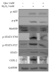
Effects of quercetin on the inflammatory response in H2O2-treated H9C2 cells. The expressed levels of phospho-Akt (Ser-473), phospho-p38 (Tyr-180/182), Mn-SOD, phospho-STAT3 (Tyr-705), phospho-STAT3 (Ser-727), COX-2, and STAT3 in H9C2 cell were detected by immunoblotting. GAPDH served as a loading control.
Figure 4
Figure 4
Effects of quercetin on ROS production in H2O2-treated H9C2 cells. The level of ROS in H9C2 cells was analyzed using a DCFH-DA assay. Values represent the mean ± standard variation for 3 independent experiments performed in triplicate and are represented as a ratio of the control. The control contains only serum-free DMEM. (***P < 0.001).
Figure 5
Figure 5
Effects of quercetin on cell apoptosis in H2O2-treated H9C2 cells. ((a), (b), and (c)) Typical dot plots of annexin V-FITC and PI are cells untreated, H2O2 treated, and quercetin pretreated followed by H2O2 treatment. The x-axis and y-axis represent the intensity of annexin V-FITC and PI, respectively. The lower left area of (a), (b), and (c) presented background staining by annexin V-FITC and PI in normal cells and apoptosis signals located in the right area. This figure is representative of 3 replicates. (d) The full lengths of DNA in H9C2 cells were detected by FACS. The x-axis shows the intensity of PI, and the y-axis shows the number of cells. (e) The levels of Bax, BCL-2, and caspase 9 in H9C2 cells were detected by immunoblotting. GAPDH served as a sample loading control.
Figure 6
Figure 6
2D-DIGE analysis of H9C2 cell proteome in response to H2O2 and quercetin treatment. H9C2 cells were lysed and arranged for a triplicate electrophoresis using pH 3 to 10 nonlinear, 24 cm IPG strips, and SDS-PAGE after treatment. 2D-DIGE image of protein sample (Cy2) is shown here. The spot numbers represent differentially expressed proteins.
Figure 7
Figure 7
Representative images of identified proteins spots and MALDI-TOF MS analysis of (a) alpha-soluble NSF attachment protein (α-SNAP); (b) elongation factor 1-alpha 1 display differentially expressed proteins among untreated, H2O2 treated, and quercetin-pretreated followed by H2O2 treatment. The differentially expressed levels of these proteins appear as 2D patterns (top images), 3D spot images (middle images), and protein abundance levels (bottom images). The PMF patterns were ((c) and (d)) from MALDI-TOF MS.
Figure 8
Figure 8
Percentage of functional distribution of differentially expressed proteins in H9C2 cell responses to H2O2 and quercetin treatment based on proteomic analysis.
Figure 9
Figure 9
Comprehensive immunofluorescence images and immunoblotting analysis of the differentially expressed proteins identified by MALDI-TOF MS. (a) The differential expression and distribution of α-SNAP in H9C2 cells responded to H2O2 and quercetin (Que) were analyzed using immunofluorescence. (b) Immunoblotting was performed to validate CDK4 and STIP1 in the H9C2 cell lysate. α-Tubulin served as a loading control. Scale bar = 20 μm.
Figure 10
Figure 10
Model illustrating how quercetin protects cardiomyocytes from ROS treatment. ROS activates Src kinase and the overexpression of α-SNAP. ROS-induced α-SNAP causes phospho-Src-FAK complexes to move from the cytosol to a nearby inner cell membrane. ROS-activated p-Src and p-p38 stimulate the phosphorylation of STAT3 at tyrosine 705 and serine 727. After p-Src kinase and p-p38 activated STAT3, the p-STAT3 dimerized to translocate into nucleus. The dimerization of p-STAT3 induces the proinflammatory response gene expression (i.e., COX-2) in oxidative stress. Ena/VASP-like protein (Evl), which participates in actin binding and homologous recombination, is upregulated in ROS-induced cells and can repair ROS-damaged DNA. Elongation factor 1-alpha 1 (Eef1a1) decreased in oxidative stress resulting in cell death. Myosin regulatory light polypeptide 9 (Myl9), profilin-1 (Pfn1), and Eef1a1 are correlated with cytoskeleton, which may induce cell deadhesion and apoptosis. ROS inhibited the expression of isopentenyl-diphosphate delta-isomerase 1 (Idi1) in cells that may block the formation of lipophilic molecular such as sterols, ubiquinones, and terpenoids. Quercetin may protect ROS-damaged cardiomyocytes via these routes (stop sign in red). The proteins (red) were altered in a H2O2-dependant manner but reverted by pretreatment with quercetin.

Similar articles

Cited by

References

    1. Takano H, Zou Y, Hasegawa H, Akazawa H, Nagai T, Komuro I. Oxidative stress-induced signal transduction pathways in cardiac myocytes: involvement of ROS in heart diseases. Antioxidants and Redox Signaling. 2003;5(6):789–794. - PubMed
    1. Chou HC, Chen YW, Lee TR, et al. Proteomics study of oxidative stress and Src kinase inhibition in H9C2 cardiomyocytes: a cell model of heart ischemia-reperfusion injury and treatment. Free Radical Biology and Medicine. 2010;49(1):96–108. - PubMed
    1. Morgan MJ, Liu ZG. Reactive oxygen species in TNFα-induced signaling and cell death. Molecules and Cells. 2010;30(1):1–12. - PMC - PubMed
    1. Rusnak F, Reiter T. Sensing electrons: protein phosphatase redox regulation. Trends in Biochemical Sciences. 2000;25(11):527–529. - PubMed
    1. Boots AW, Haenen GRMM, Bast A. Health effects of quercetin: from antioxidant to nutraceutical. European Journal of Pharmacology. 2008;585(2-3):325–337. - PubMed