Effects of mechano-electric feedback on scroll wave stability in human ventricular fibrillation
- PMID: 23573245
- PMCID: PMC3616032
- DOI: 10.1371/journal.pone.0060287
Effects of mechano-electric feedback on scroll wave stability in human ventricular fibrillation
Abstract
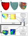
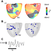
Recruitment of stretch-activated channels, one of the mechanisms of mechano-electric feedback, has been shown to influence the stability of scroll waves, the waves that underlie reentrant arrhythmias. However, a comprehensive study to examine the effects of recruitment of stretch-activated channels with different reversal potentials and conductances on scroll wave stability has not been undertaken; the mechanisms by which stretch-activated channel opening alters scroll wave stability are also not well understood. The goals of this study were to test the hypothesis that recruitment of stretch-activated channels affects scroll wave stability differently depending on stretch-activated channel reversal potential and channel conductance, and to uncover the relevant mechanisms underlying the observed behaviors. We developed a strongly-coupled model of human ventricular electromechanics that incorporated human ventricular geometry and fiber and sheet orientation reconstructed from MR and diffusion tensor MR images. Since a wide variety of reversal potentials and channel conductances have been reported for stretch-activated channels, two reversal potentials, -60 mV and -10 mV, and a range of channel conductances (0 to 0.07 mS/µF) were implemented. Opening of stretch-activated channels with a reversal potential of -60 mV diminished scroll wave breakup for all values of conductances by flattening heterogeneously the action potential duration restitution curve. Opening of stretch-activated channels with a reversal potential of -10 mV inhibited partially scroll wave breakup at low conductance values (from 0.02 to 0.04 mS/µF) by flattening heterogeneously the conduction velocity restitution relation. For large conductance values (>0.05 mS/µF), recruitment of stretch-activated channels with a reversal potential of -10 mV did not reduce the likelihood of scroll wave breakup because Na channel inactivation in regions of large stretch led to conduction block, which counteracted the increased scroll wave stability due to an overall flatter conduction velocity restitution.
Conflict of interest statement
Figures


Similar articles
-
Transmural dispersion of repolarization determines scroll wave behavior during ventricular tachyarrhythmias.Circ J. 2011;75(1):80-8. doi: 10.1253/circj.cj-10-0071. Epub 2010 Nov 16. Circ J. 2011. PMID: 21099125
-
Scroll wave dynamics in a three-dimensional cardiac tissue model: roles of restitution, thickness, and fiber rotation.Biophys J. 2000 Jun;78(6):2761-75. doi: 10.1016/S0006-3495(00)76821-4. Biophys J. 2000. PMID: 10827961 Free PMC article.
-
Voltage-induced slow activation and deactivation of mechanosensitive channels in Xenopus oocytes.J Physiol. 1997 Dec 15;505 ( Pt 3)(Pt 3):551-69. doi: 10.1111/j.1469-7793.1997.551ba.x. J Physiol. 1997. PMID: 9457635 Free PMC article.
-
Cardiac mechano-electric feedback and electrical restitution in humans.Prog Biophys Mol Biol. 2008 Jun-Jul;97(2-3):452-60. doi: 10.1016/j.pbiomolbio.2008.02.021. Epub 2008 Feb 16. Prog Biophys Mol Biol. 2008. PMID: 18407323 Review.
-
Molecular mechanisms and global dynamics of fibrillation: an integrative approach to the underlying basis of vortex-like reentry.J Theor Biol. 2004 Oct 21;230(4):475-87. doi: 10.1016/j.jtbi.2004.02.024. J Theor Biol. 2004. PMID: 15363670 Review.
Cited by
-
Mechano-electric feedback effects in a three-dimensional (3D) model of the contracting cardiac ventricle.PLoS One. 2018 Jan 17;13(1):e0191238. doi: 10.1371/journal.pone.0191238. eCollection 2018. PLoS One. 2018. PMID: 29342222 Free PMC article.
-
A Mathematical Model of Neonatal Rat Atrial Monolayers with Constitutively Active Acetylcholine-Mediated K+ Current.PLoS Comput Biol. 2016 Jun 22;12(6):e1004946. doi: 10.1371/journal.pcbi.1004946. eCollection 2016 Jun. PLoS Comput Biol. 2016. PMID: 27332890 Free PMC article.
-
Methodology for image-based reconstruction of ventricular geometry for patient-specific modeling of cardiac electrophysiology.Prog Biophys Mol Biol. 2014 Aug;115(2-3):226-34. doi: 10.1016/j.pbiomolbio.2014.08.009. Epub 2014 Aug 19. Prog Biophys Mol Biol. 2014. PMID: 25148771 Free PMC article.
-
Evaluating functional capacity, and mortality effects in the presence of atrial electromechanical conduction delay in patients with systolic heart failure.Anatol J Cardiol. 2016 Aug;16(8):579-586. doi: 10.5152/AnatolJCardiol.2015.6445. Epub 2015 Oct 21. Anatol J Cardiol. 2016. PMID: 27004707 Free PMC article.
-
Chaos control in cardiac dynamics: terminating chaotic states with local minima pacing.Front Netw Physiol. 2024 Jul 3;4:1401661. doi: 10.3389/fnetp.2024.1401661. eCollection 2024. Front Netw Physiol. 2024. PMID: 39022296 Free PMC article.
References
-
- Kohl P, Hunter P, Noble D (1999) Stretch-induced changes in heart rate and rhythm: clinical observations, experiments and mathematical models. Prog Biophys Mol Biol 71: 91–138. - PubMed
-
- Kohl P, Ravens U (2003) Cardiac mechano-electric feedback: past, present, and prospect. Prog Biophys Mol Biol 82: 3–9. - PubMed
-
- Miragoli M, Gaudesius G, Rohr S (2006) Electrotonic modulation of cardiac impulse conduction by myofibroblasts. Circ Res 98: 801–810. - PubMed
-
- Hu H, Sachs F (1997) Stretch-activated ion channels in the heart. J Mol Cell Cardiol 29: 1511–1523. - PubMed
Publication types
MeSH terms
Grants and funding
LinkOut - more resources
Full Text Sources
Other Literature Sources
Miscellaneous

