. 2013;8(4):e60445.
doi: 10.1371/journal.pone.0060445. Epub 2013 Apr 3.

Crystal structure of KLHL3 in complex with Cullin3

Affiliations

Crystal structure of KLHL3 in complex with Cullin3

Alan X Ji et al. PLoS One. 2013.

Abstract

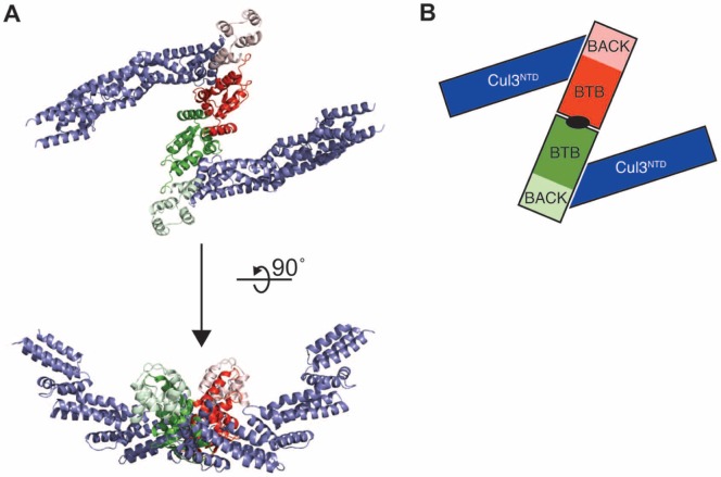
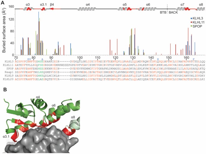
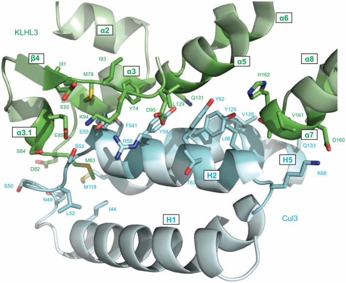
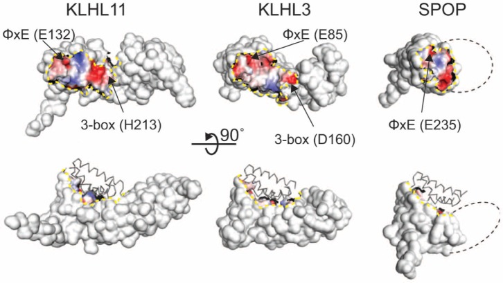
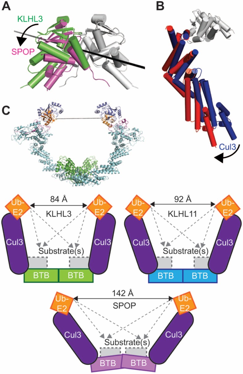
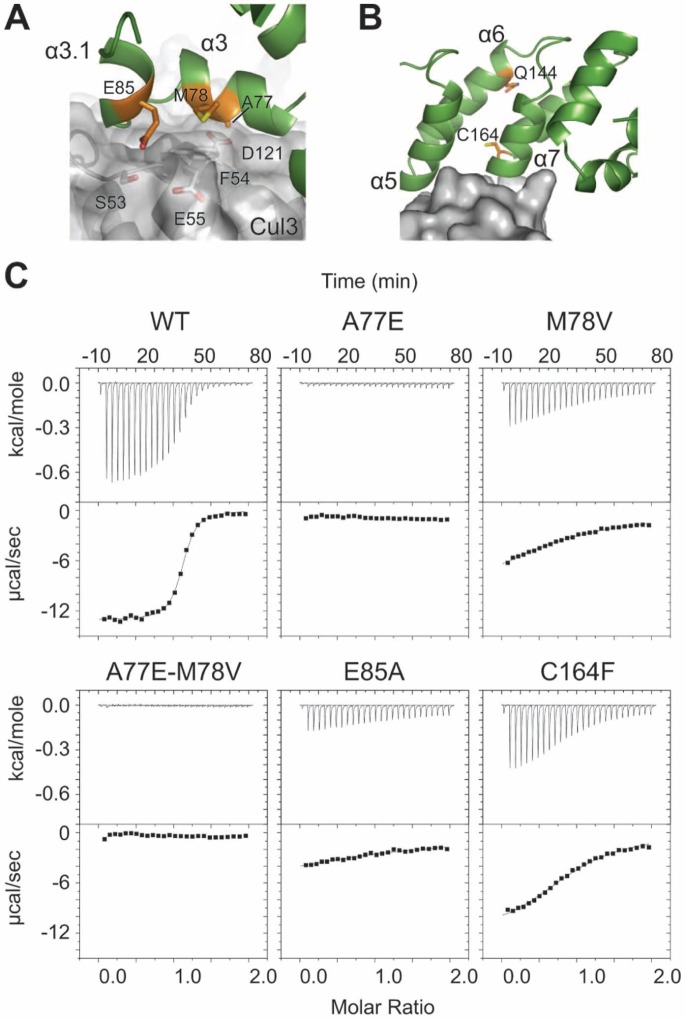
KLHL3 is a BTB-BACK-Kelch family protein that serves as a substrate adapter in Cullin3 (Cul3) E3 ubiquitin ligase complexes. KLHL3 is highly expressed in distal nephron tubules where it is involved in the regulation of electrolyte homeostasis and blood pressure. Mutations in KLHL3 have been identified in patients with inherited hypertension disorders, and several of the disease-associated mutations are located in the presumed Cul3 binding region. Here, we report the crystal structure of a complex between the KLHL3 BTB-BACK domain dimer and two copies of an N terminal fragment of Cul3. We use isothermal titration calorimetry to directly demonstrate that several of the disease mutations in the KLHL3 BTB-BACK domains disrupt the association with Cul3. Both the BTB and BACK domains contribute to the Cul3 interaction surface, and an extended model of the dimeric CRL3 complex places the two E2 binding sites in a suprafacial arrangement with respect to the presumed substrate-binding sites.


Conflict of interest statement

Competing Interests: The authors have declared that no competing interests exist.

Figures

Figure 1
Figure 1. Crystal structure of the 2∶2 KLHL3BTB-BACK/Cul3NTD complex.
(A) The KLHL3BTB-BACK homodimer (green and red) binds to two Cul3NTD chains (blue). The KLHL3 BTB domains are in bold colors, and the BACK domains are in lighter colors. (B) Schematic of the 2∶2 complex. The view is along the BTB dimerization axis, indicated in black.
Figure 2
Figure 2. The KLHL3/Cul3 interface.
(A) Residue-based buried surface area for KLHL3 (PDB ID 4HXI), KLHL11 (PDB ID 4AP2), and SPOP (PDB ID 4EOZ) when bound to Cul3. The multiple sequence alignment of Cul3 interacting proteins is colored by conservation, with the φ-x-E motif residues in green. Mutations identified in PHAII are indicated in blue above the KLHL3 sequence. “X” indicates a stop codon. (B) KLHL3 is shown in green ribbons with residue positions in contact with Cul3 highlighted in red. Cul3 is shown as a grey surface. The KLHL3 BACK domain is colored light green.
Figure 3
Figure 3. Details of the KLHL3BTB-BACK/Cul3NTD interface.
The side chains of the residues at the protein-protein interface are indicated.
Figure 4
Figure 4. Cul3 binding surfaces of BTB-BACK proteins.
The solvent-accessible surfaces of KLHL3BTB-BACK, KLHL11BTB-BACK and SPOPBTB are shown, with the Cul3-contacting region colored by the electrostatic potential, as indicated. A dashed yellow line delineates the Cul3-contacting region. The dashed black line indicates the approximate region where the BACK domain would be found in the SPOP structure. The three proteins are in similar orientations in the two views. Cul3 is shown in Cα trace in the lower set of structures.
Figure 5
Figure 5. Differences in the quaternary structures of BTB/Cul3 complexes.
(A) Comparison of the dimerization interfaces of the KLHL3 and SPOP BTB domains. A single BTB chain from KLHL3 and SPOP was superposed (shown in white and grey), resulting in a misalignment of the partner BTB chains. The second chain in the KLHL3 BTB dimer is shown in green, and the second chain of the SPOP BTB dimer is shown in magenta. The axis of rotation is shown as a black line and is approximately normal to the BTB dimerization axis. (B) Comparison of Cul3 chains from KLHL3 (blue) and SPOP (red) after aligning BTB domains (C) Model and schematics of fully assembled BTB/Cul3/E2/Ubiquitin complexes. The distances between the E2 ubiquitin conjugation sites are shown as solid arrows, and the dashed grey arrows illustrate distances to substrate binding locations. They grey regions indicate the substrate-binding Kelch domains in KLHL3 and KLHL11, and the MATH domain in SPOP.
Figure 6
Figure 6. KLHL3 BTB-BACK domain mutations in PHAII.
KLHL3 is shown as a green ribbon and Cul3 is shown as a transparent grey surface. Relevant PHAII mutations are colored orange. (A) KLHL3 residues A77, M78 and E85 are in direct contact with Cul3. (B) KLHL3 residue C164 is near, but not in direct contact with Cul3. Q144 indicates the location of the PHAII nonsense mutation. (C) ITC data and binding isotherms for the KLHL3BTB-BACK/Cul3NTD interaction for wild-type and KLHL3 PHAII mutants.

Similar articles

Cited by

References

    1. Petroski MD, Deshaies RJ (2005) Function and regulation of cullin-RING ubiquitin ligases. Nat Rev Mol Cell Biol 6: 9–20. - PubMed
    1. Sarikas A, Hartmann T, Pan ZQ (2011) The cullin protein family. Genome Biol 12: 220. - PMC - PubMed
    1. Furukawa M, He YJ, Borchers C, Xiong Y (2003) Targeting of protein ubiquitination by BTB-Cullin 3-Roc1 ubiquitin ligases. Nat Cell Biol 5: 1001–1007. - PubMed
    1. Zheng N, Schulman BA, Song L, Miller JJ, Jeffrey PD, et al. (2002) Structure of the Cul1-Rbx1-Skp1-F boxSkp2 SCF ubiquitin ligase complex. Nature 416: 703–709. - PubMed
    1. Angers S, Li T, Yi X, MacCoss MJ, Moon RT, et al. (2006) Molecular architecture and assembly of the DDB1-CUL4A ubiquitin ligase machinery. Nature 443: 590–593. - PubMed

Publication types

MeSH terms