. 2013;8(4):e60784.
doi: 10.1371/journal.pone.0060784. Epub 2013 Apr 1.

Tacrolimus inhibits NF-κB activation in peripheral human T cells

Affiliations

Tacrolimus inhibits NF-κB activation in peripheral human T cells

Ramin Vafadari et al. PLoS One. 2013.

Abstract

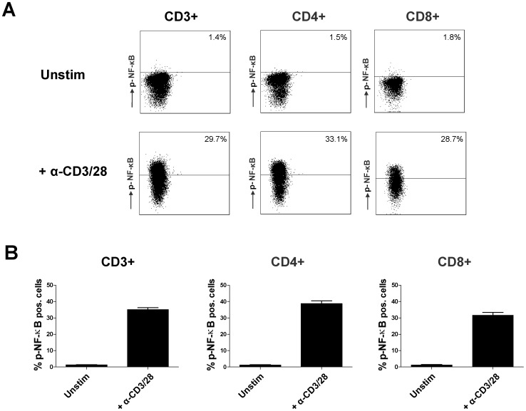
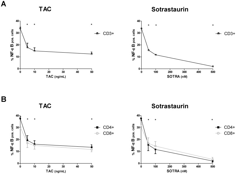
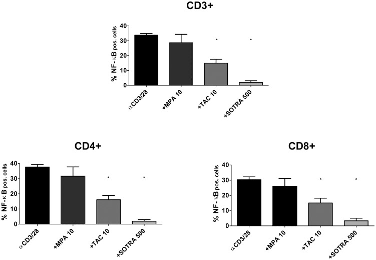
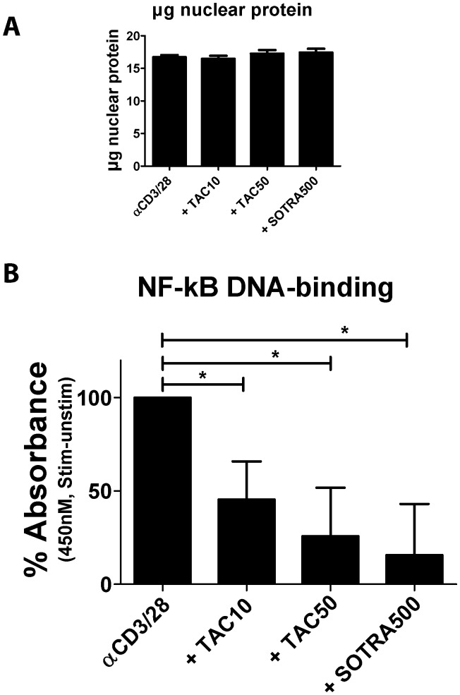
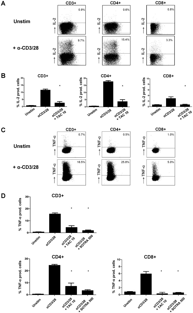
The calcineurin inhibitor, tacrolimus (TAC), inhibits the protein phosphatase activity of calcineurin, leading to suppression of the nuclear translocation of NFAT and inhibition of T cell activation. Apart from NFAT also the transcription factor NF-κB plays a key functional role in T cell activation. Therefore, blockade of the NF-κB activation cascade by immunosuppressive drugs prevents immune activation. Here we studied whether TAC blocks NF-κB activation in peripheral human T cells. After anti-CD3/CD28-activation of T cells from healthy volunteers, NF-κB (p65) phosphorylation was measured by flow cytometry in CD3+ T cells, CD4+ helper T cells and CD8+ cytotoxic T cells in the absence and presence of TAC 10 ng/mL, sotrastaurin 500 nM (positive control) and mycophenolic acid 10 µg/mL (negative control; n = 6). NF-κB transcriptional activity was measured by ELISA and intracellular TNFα protein, a downstream target, was measured by flow cytometry to assess the functional consequences of NF-κB blockade. Anti-CD3/28-activation induced NF-κB phosphorylation in CD3+ T cells, CD4+ T cells and CD8+ T cells by 34% (mean), 38% and 30% resp. (p<0.01). Sotrastaurin inhibited NF-κB activation in the respective T cell subsets by 93%, 95% and 86% (p<0.01 vs. no drug), while mycophenolic acid did not affect this activation pathway. Surprisingly, TAC also inhibited NF-κB phosphorylation, by 55% (p<0.01) in CD3+ T cells, by 56% (p<0.01) in CD4+ T cells and by 51% in CD8+ T cells (p<0.01). In addition, TAC suppressed NF-κB DNA binding capacity by 55% (p<0.05) in CD3+ T cells and TNFα protein expression was inhibited in CD3+ T cells, CD4+ T cells and CD8+ T cells by 76%, 71% and 93% resp. (p<0.01 vs. no drug), confirming impaired NF-κB signaling. This study shows the suppressive effect of TAC on NF-κB signaling in peripheral human T cell subsets, measured at three specific positions in the NF-κB activation cascade.


Conflict of interest statement

Competing Interests: The authors have declared that no competing interests exist.

Figures

Figure 1
Figure 1. Facs analysis of NF-κB phosphorylation in T cells.
The total CD3+ T cell population was acquired by MACS isolation of PBMC from healthy volunteers. A) Example scatter dot plots of an unstimulated sample (above) and an anti-CD3/CD28 stimulated sample to illustrate the gating strategy for analysis of percentages CD3+, CD4+ and CD8+ T cells which express phosphorylated NF-κB. Both samples were stained with a monoclonal antibody against phosphorylated NF-κB p65. B) The average percentages of cells with elevated NF-κB phosphorylation in unstimulated and stimulated samples without drug are depicted as mean ± SEM of 6 independent experiments.
Figure 2
Figure 2. NF-κB phosphorylation inhibited at different TAC-concentrations.
MACS isolated CD3+ T cells from healthy volunteers were stimulated by anti-CD3/CD28 in the presence of TAC 0, 5, 10, 50 ng/mL or sotrastaurin 50, 100 and 500 nM. Inhibition of NF-κB phosphorylation is shown for CD3+ T cells (above), CD4+ T cells and CD8+ T cells (below; mean ± SEM of 6 independent experiments).
Figure 3
Figure 3. NF-κB phosphorylation inhibited by immunosuppressive drugs.
The effect of the immunosuppressive drugs mycophenolic acid 10 µg/mL, TAC 10 ng/mL and sotrastaurin 500 nM on NF-κB p65 phosphorylation in peripheral T cells from healthy volunteers is depicted. Mycophenolic acid did not influence NF-κB phosphorylation (p>0.05), while the positive control sotrastaurin inhibited phosphorylation in the CD3+, CD4+ and CD8+ T cell subsets (p<0.01 compared to no drug). TAC 10 ng/mL also suppressed phosphorylation in the T cell subsets (p<0.01; mean ± SEM of 6 independent experiments).
Figure 4
Figure 4. TAC inhibits NF-κB DNA-binding capacity in human T cells.
MACS isolated CD3+ T cells (3.106) from three healthy volunteers were stimulated in the presence of TAC 0, 10, 50 ng/mL or sotrastaurin 500 nM. A) The amount of nuclear protein extracted from the cells was comparable in the samples and at the different tested conditions. B) The DNA binding capacity of NF-κB in the nuclear fractions was inhibited by both TAC and the positive control sotrastaurin. Each bar represents the anti-CD3/CD28-stimulated NF-κB DNA binding capacity minus that of the unstimulated sample (mean ± SEM of 3 independent experiments).
Figure 5
Figure 5. TAC induces inhibition of NF-κB dependent cytokine production.
CD3+ T cells were acquired by MACS isolation of PBMC from healthy volunteers. Cells were anti-CD3/CD28 stimulated for 24 hours in the presence of TAC 0 and 10 ng/mL; and sotrastaurin 500 nM. A) Dot plots showing CD3+, CD4+ and CD8+ T cells with intracellular IL-2 production in an unstimulated and a stimulated sample. Both sample-types were stained for IL-2 protein expression. B) The percentage of IL-2 producing anti-CD3/CD28 activated T cells was fully abrogated by tacrolimus 10 ng/mL (p<0.01; mean ± SEM of 4 independent experiments). C) Dot plots showing CD3+, CD4+ and CD8+ T cells with intracellular TNFα production in an unstimulated and a stimulated sample. Both sample-types were stained with a monoclonal antibody against TNFα. D) The average percentages of TNFα producing T cells in CD3+, CD4+ and CD8+ primary T cells are depicted. TAC 10 ng/mL and sotrastaurin 500nM both inhibited the induced TNFα expression in the T cell subsets (p<0.01; mean ± SEM of 4 independent experiments).

Similar articles

Cited by

References

    1. Meier-Kriesche HU, Li S, Gruessner RW, Fung JJ, Bustami RT, et al. (2006) Immunosuppression: evolution in practice and trends, 1994-2004. Am J Transplant 6: 1111–1131. - PubMed
    1. Cecka JM (2005) The OPTN/UNOS Renal Transplant Registry. Clin Transpl: 1-16. - PubMed
    1. Tang IY, Meier-Kriesche HU, Kaplan B (2007) Immunosuppressive strategies to improve outcomes of kidney transplantation. Semin Nephrol 27: 377–392. - PubMed
    1. Ekberg H, Tedesco-Silva H, Demirbas A, Vitko S, Nashan B, et al. (2007) Reduced exposure to calcineurin inhibitors in renal transplantation. N Engl J Med 357: 2562–2575. - PubMed
    1. Ekberg H, Grinyo J, Nashan B, Vanrenterghem Y, Vincenti F, et al. (2007) Cyclosporine sparing with mycophenolate mofetil, daclizumab and corticosteroids in renal allograft recipients: the CAESAR Study. Am J Transplant 7: 560–570. - PubMed