. 2013;8(4):e61027.
doi: 10.1371/journal.pone.0061027. Epub 2013 Apr 3.

WWP1 E3 ligase targets LATS1 for ubiquitin-mediated degradation in breast cancer cells

Affiliations

WWP1 E3 ligase targets LATS1 for ubiquitin-mediated degradation in breast cancer cells

Benjamin Yeung et al. PLoS One. 2013.

Abstract

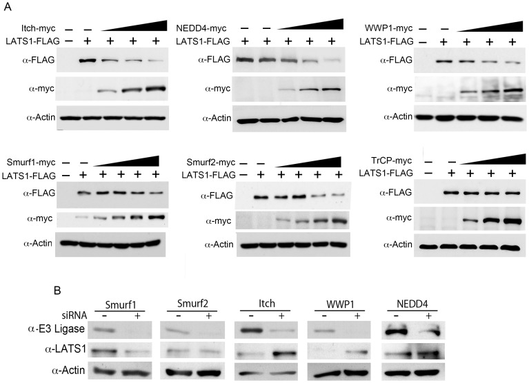
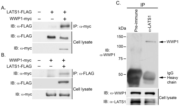
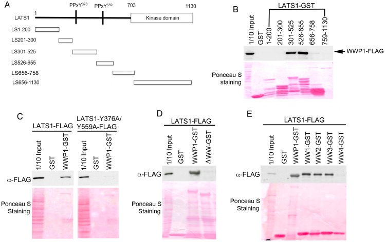
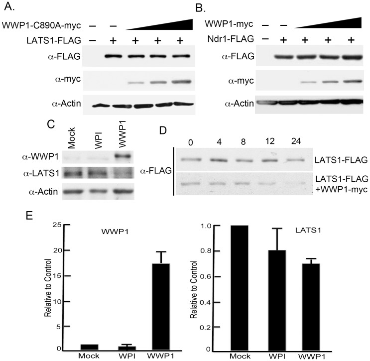
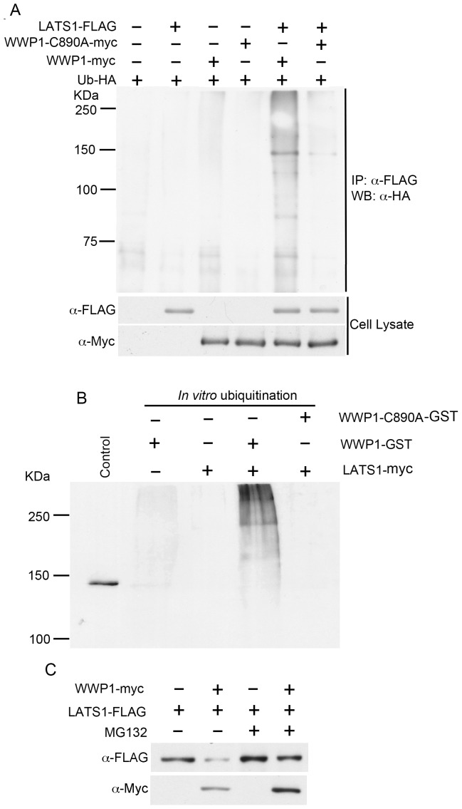
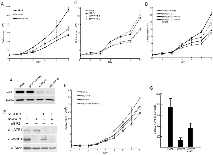
The Large Tumor Suppressor 1 (LATS1) is a serine/threonine kinase and tumor suppressor found down-regulated in various human cancers. LATS1 has recently been identified as a central player of the emerging Hippo signaling pathway, which plays important roles in organ size control, tumorigenesis, and stem cell differentiation and renewal, etc. Although mounting evidence supports a role of LATS1 in tumor suppression and tumorigenesis, how LATS1 is regulated at the molecular level is not fully understood. Recently several positive regulators of LATS1 (Mst1/2, MOB1, Kibra, etc) have been identified but how LATS1 is negatively regulated is still largely unknown. We have recently identified Itch, a member of the NEDD4-like family E3 ubiquitin ligases, as a novel negative regulator of LATS1. However, whether other ubiquitin ligases modulate LATS1 stability and function is unclear. By screening many E3 ligases of the NEDD4-like family using over-expression and short-interference RNA knockdown approaches, we have identified WWP1 E3 ligase as another novel negative regulator of LATS1. We have provided in vitro and in vivo evidence that WWP1 is essential for LATS1 stability and negatively regulate LATS1 by promoting LATS1 degradation through polyubiquitination and the 26S proteasome pathway. Importantly, we also showed that degradation of LATS1 is critical in mediating WWP1-induced increased cell proliferation in breast cancer cells. Since WWP1 is an oncogene and LATS1 is a tumor suppressor gene in breast cancer, our studies provide a promising therapeutic strategy in which developed drugs targeting WWP1 cause activation of LATS1 in suppressing breast cancer cell growth.


Conflict of interest statement

Competing Interests: The authors have declared that no competing interests exist.

Figures

Figure 1
Figure 1. Effect of NEDD4-like family E3 ubiquitin ligases on LATS1 stability.
A) Dose-dependent degradation of LATS1 by E3 ubiquitin ligases. LATS1-FLAG plasmids were transfected alone (0.2 µg) or together with increasing amounts [0.05, 0.1, 0.2, 0.4 µg] of WWP1 plasmids into COS7 cells. The levels of LATS1-FLAG and WWP1-myc were detected by western blotting using anti-FLAG and anti-Myc antibodies, respectively. B) Knockdown of NEDD4-like family E3 ubiquitin ligases on LATS1 stability. About 100 nM of siRNA targeting scrambled RNA (negative control) or each E3 ligase was transfected into MCF7 or T47D cells in a 6-well plate. Two days after transfection, proteins were extracted and subjected to western blot analysis of each E3 ligase, LATS1, and β-actin (internal loading control).
Figure 2
Figure 2. Interaction of LATS1 and WWP1 in vivo.
A, B) Co-IP of ectopically expressed LATS1 and WWP1 in vivo. COS7 cell lysates expressing either LATS1-FLAG (A) or WWP1-myc (B) alone or together with WWP1-myc (A) or LATS1-FLAG (B) were immunoprecipitated with anti-Myc (A) or anti-FLAG (B) antibody, followed by western blotting with anti-FLAG antibody (A) or anti-Myc (B) antibody, respectively. Expression of LATS1 levels in cell lysate was also examined by western blot (A, B). C) Co-IP of endogenous LATS1 and WWP1 in vivo. About 1 mg of cell lysates from HEK293T were immunoprecipitated with either control pre-immune serum or rabbit anti-LATS1 (Y03) polyclonal antibody, followed by western blotting with anti-WWP1 antibody. Expression of endogenous WWP1 and LATS1 is also shown.
Figure 3
Figure 3. Interaction of LATS1 and WWP1 in vitro.
A) Diagram of LATS1 deletion. B-E) GST pulldown analysis of functional domains responsible for the interaction of LATS1 and WWP1. About 100 µg of protein lysates from COS7 cells expressing FLAG-tagged LATS1 or its mutants (C–E), or WWP1 (B) was mixed with 10 µg of GST (control) or GST fusion proteins (LATS1 deletions or WWP1 mutants) on beads. The beads were washed and the binding proteins were eluted and subjected to western blot using anti-FLAG antibody. 1/10 input (10 µg) represents 1/10 of protein lysate (100 µg) used for GST pulldown. Ponceau S was used to stain fusion proteins on the membrane. B) Functional domains of LATS1 interacting with WWP1. Lysate: WWP1-FLAG; LATS1 deletion GST fusion proteins. C) Interaction of LATS1-PPxY mutants with WWP1 in vitro. Lysate: LATS1-FLAG (wild-type) or LATS1 with double PPxY mutation (LATS1-Y376A/Y559A-FLAG). D) Functional domains of WWP1 interacting with LATS1 in vitro. ΔWW-GST: WWP1 lacking all of 4 WW domains. E) Interaction of LATS1 with each WW domain of WWP1 in vitro. WW1-4 GST: first to fourth WW domain GST fusion protein.
Figure 4
Figure 4. Regulation of LATS1 protein stability by WWP1.
A) Ligase-dependent degradation of LATS1 by WWP1. Increasing amounts of ligase-dead WWP1 (WWP1-C890A-myc) was co-transfected with equal amounts of LATS1 plasmids. B) Inability of WWP1 to degrade LATS1 homolog Ndr1 lacking PPxY motifs. C) Degradation of endogenous LATS1 by WWP1 in MCF10A mammary cells. MCF10A cells were mock infected or infected with WPI (vector) or WWP1-WPI lentivirus. After establishment of stable lines, endogenous WWP1, LATS1, or β-actin was detected by western blot. D) Measurement of WWP1 half-life by CHX chase assays. LATS1-FLAG alone or together with WWP1-myc were transfected into COS7 cells, followed by treatment with CHX to inhibit protein synthesis. At the indicated time, cells are harvested and analyzed for LATS1 level using anti-FLAG antibody. E) Expression of LATS1 mRNA after WWP1 over-expression. About 0.1 µg of RNAs was used for one-step qRT-PCR (Invitrogen) analysis using rRNA as controls. Values represent mean and standard deviation of fold increase of mRNA levels in WPI or WWP1-expressing MCF10A cells relative to those in mock-infected MCF10A cells.
Figure 5
Figure 5. WWP1 promotes LATS1 degradation by polyubiquitination and the proteasome pathway.
A) Ubiquitination of LATS1 by WWP1 in vivo. Ubiquitin-HA and different combination of WWP1-myc, WWP1-ligase-dead mutant (WWP1-C890A-myc) and LATS1-FLAG were transfected into HEK293T cells. Ubiquitinated LATS1 was detected by immunoprecipitation of LATS1 with anti-FLAG antibody, followed by detection of ubiquitin using anti-HA antibody. B) Ubiquitination of LATS1 by WWP1 in vitro. Immunoprecipitated LATS1-myc or LATS1-Y376A/Y559A-myc on beads was used as a substrate in an ubiquitination assay with a ligase buffer containing E1, E2, Ubiquitin-FLAG, ATP, and WWP1-GST or WWP1-C890A-GST. After the reaction, beads containing LATS1-myc were washed extensively with modified RIPA buffer, followed by western blot analysis using anti-FLAG antibody. Cell lysate expressing FLAG-tagged LATS1 was used as positive control (lane 1). C) Inhibition of WWP1-mediated LATS1 degradation by proteasome inhibitor MG132. COS7 cells transfected with either LATS1-FLAG alone or together with WWP1-myc were treated with either DMSO (control) or proteasome inhibitor (MG132), followed by examination of LATS1 protein levels by western blot.
Figure 6
Figure 6. LATS1 mediates the effect of WWP1 on cell proliferation.
A) Increased cell proliferation after WWP1 over-expression in MCF10A cells. Triplicate of 2×10⁴ infected MCF10A cells (mock) or MCF10A cells expressing WPI (vector) or WWP1-WPI were plated into each well of 24-well plate, followed by cell count for 5 days. The mean and standard deviation (SD) of three samples for each day are shown. B, C) Knockdown of WWP1 reduces cell proliferation assay. MCF7 cells were mock-infected or stably infected with lentivirus expressing pGIPZ (vector) or two shWWP1 shRNAs (shWWP1-1 and shWWP1-2). The levels of WWP1 and β-actin were examined by western blot (B). Cell proliferation analysis for these lines was performed for 6 days. The mean and standard deviation of three samples for each day are shown (C). D) Rescue of shWWP1-induced reduced cell proliferation by wild-type rather than ligase dead (WWP1-C890A). Cell proliferation was performed as described in (B). E, F) Knockdown of LATS1 rescues shWWP1-induced reduced cell proliferation. MCF7 cells were infected with lentivirus expressing pGIPZ (vector), shLATS1, shWWP1-1, and shWWP1-1/shLATS1. The levels of LATS1, WWP1, and β-actin were examined by western blot (E). Cell proliferation analysis was carried out for 8 days. The mean and standard deviation of three samples for each day are shown (F). G) Colony forming assay. About 1000 cells were plated in each of 100 mm plate, followed by incubation the cells at 37°C for 10 days. The mean and standard deviation of colony numbers from three plates for each cell line are shown.

Similar articles

Cited by

References

    1. Visser S, Yang X (2010) LATS tumor suppressor: A new governor of cellular homeostasis. Cell Cycle 9: 3892–3903. - PubMed
    1. Yang X, Yu K, Hao Y, Li DM, Stewart R, et al. (2004) LATS1 tumour suppressor affects cytokinesis by inhibiting LIMK1. Nature Cell Biology 6: 609–617. - PubMed
    1. Yang X, Li DM, Chen W, Xu T (2001) Human homologue of drosophila lats, LATS1, negatively regulate growth by inducing G(2)/M arrest or apoptosis. Oncogene 20: 6516–23. - PubMed
    1. Lai D, Ho KC, Hao Y, Yang X (2011) Taxol resistance in breast cancer cells is mediated by the hippo pathway component TAZ and its downstream transcriptional targets Cyr61 and CTGF. Cancer Research 71: 2728–2738. - PubMed
    1. Tremblay AM, Camargo FD (2012) Hippo signaling in mammalian stem cells. Seminars in Cell & Developmental Biology 23: 818–826. - PubMed

Publication types

MeSH terms

Substances