. 2013;8(4):e61267.
doi: 10.1371/journal.pone.0061267. Epub 2013 Apr 3.

EGFR-targeted granzyme B expressed in NK cells enhances natural cytotoxicity and mediates specific killing of tumor cells

Affiliations

EGFR-targeted granzyme B expressed in NK cells enhances natural cytotoxicity and mediates specific killing of tumor cells

Pranav Oberoi et al. PLoS One. 2013.

Abstract

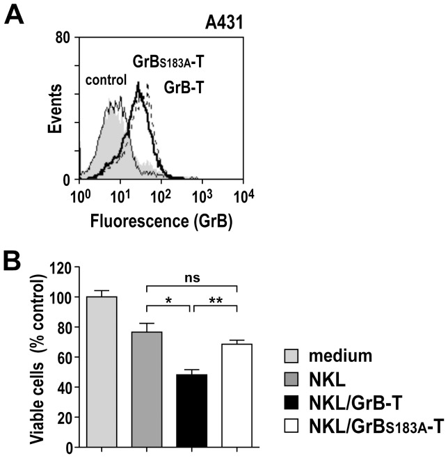
Natural killer (NK) cells are highly specialized effectors of the innate immune system that hold promise for adoptive cancer immunotherapy. Their cell killing activity is primarily mediated by the pro-apoptotic serine protease granzyme B (GrB), which enters targets cells with the help of the pore-forming protein perforin. We investigated expression of a chimeric GrB fusion protein in NK cells as a means to augment their antitumoral activity. For selective targeting to tumor cells, we fused the epidermal growth factor receptor (EGFR) peptide ligand transforming growth factor α (TGFα) to human pre-pro-GrB. Established human NKL natural killer cells transduced with a lentiviral vector expressed this GrB-TGFα (GrB-T) molecule in amounts comparable to endogenous wildtype GrB. Activation of the genetically modified NK cells by cognate target cells resulted in the release of GrB-T together with endogenous granzymes and perforin, which augmented the effector cells' natural cytotoxicity against NK-sensitive tumor cells. Likewise, GrB-T was released into the extracellular space upon induction of degranulation with PMA and ionomycin. Secreted GrB-T fusion protein displayed specific binding to EGFR-overexpressing tumor cells, enzymatic activity, and selective target cell killing in the presence of an endosomolytic activity. Our data demonstrate that ectopic expression of a targeted GrB fusion protein in NK cells is feasible and can enhance antitumoral activity of the effector cells.


Conflict of interest statement

Competing Interests: The authors have declared that no competing interests exist.

Figures

Figure 1
Figure 1. Expression of the granzyme B-TGFα fusion protein GrB-T in NK cells.
(A) Schematic representation of the lentiviral transfer vector pS-GrB-T-IEW that encodes under the control of the Spleen Focus Forming Virus promoter (SFFV) a fusion of human GrB with TGFα, followed by an internal ribosome entry site (IRES) and cDNA encoding enhanced green fluorescent protein (EGFP) as a marker. SP, GrB signal peptide; DP, GrB activation dipeptide; GrB21–247, mature form of GrB; L, flexible linker; M, Myc tag; H, hexa-histidine tag. The similar transfer vector pS-GrBS183A-T-IEW encodes enzymatically inactive mutant GrBS183A fused to TGFα (not shown). After transduction with S-GrB-T-IEW or S-GrBS183A-T-IEW vector particles, EGFP-expressing NKL/GrB-T and NKL/GrBS183A-T cells were enriched by flow cytometric cell sorting, and analyzed for GrB-T expression. (B) GrB-T mRNA expression in NKL/GrB-T and NKL/GrBS183A-T cells was verified by semi-quantitative RT-PCR. (C) Expression of GrB-T proteins in NKL/GrB-T and NKL/GrBS183A-T cells was investigated by immunoblot analysis of cell lysates with GrB-specific antibody. γ-Tubulin was analyzed as a loading control. (D) Expression of GrB-T proteins in NKL/GrB-T and NKL/GrBS183A-T cells was confirmed by intracellular staining with Myc-tag-specific antibody and flow cytometry (open areas). In all experiments parental NKL cells served as controls.
Figure 2
Figure 2. Natural cytotoxicity of NKL/GrB-T and NKL/GrBS183A-T cells.
(A) Total levels of GrB and perforin expressed by parental NKL (dark gray areas), NKL/GrB-T (bold lines) and NKL/GrBS183A-T cells (dotted lines) was analyzed by intracellular staining with GrB-specific antibody (left) or perforin-specific antibody (right) and flow cytometry. NKL cells incubated with isotype-matched antibodies served as controls (light gray areas). (B) Cytotoxicity of NKL/GrB-T (filled squares) and NKL/GrBS183A-T cells (open squares) towards C1R-neo and Jurkat cells was determined in FACS-based cytotoxicity assays at different effector to target ratios (E/T). Parental NKL cells (filled circles) were included for comparison. Dependence of target cell killing on the release of granular proteins was confirmed by incubating C1R-neo target cells with NKL effector cells in the presence of 2 mM of the Ca2+ chelator EGTA. Representative data of one of three independent experiments are shown. Absence of EGFR expression on the surface of C1R-neo and Jurkat cells was confirmed by flow cytometry with EGFR-specific antibody (open areas). Cells treated only with secondary antibody served as controls (shaded areas).
Figure 3
Figure 3. Cytotoxicity of NKL/GrB-T and NKL/GrBS183A-T cells towards EGFR-expressing tumor cells.
(A) Expression of EGFR on the surface of MDA-MB468 breast carcinoma and A431 squamous cell carcinoma cells was determined by flow cytometry with EGFR-specific antibody (open areas). Cells treated only with secondary antibody served as controls (shaded areas). (B) Cytotoxicity of NKL/GrB-T (filled squares) and NKL/GrBS183A-T cells (open squares) towards MDA-MB468 and A431 cells was determined in FACS-based cytotoxicity assays at different E/T ratios. Parental NKL cells (filled circles) were included for comparison.
Figure 4
Figure 4. Conjugate formation and redistribution of cytotoxic granules upon target cell contact.
(A) Parental NKL and NKL/GrB-T cells were incubated with C1R-neo or MDA-MB468 target cells at an E/T ratio of 1∶1 for 1 h at 37°C, stained with perforin-specific antibody as indicated (red), and analyzed by confocal laser scanning microscopy. As a control, NK cells were incubated without target cells. Maximum projection of a z-stack series is shown in each case. N: NK cell; T: target cell. (B) To assess activation of NK cells upon target cell contact, parental NKL and NKL/GrB-T cells were incubated with C1R-neo (upper panel) or MDA-MB468 target cells (lower panel) at an E/T ratio of 1∶1 for 5 h at 37°C, before analysis of cell surface-associated degranulation marker CD107a by flow cytometry (open areas). NKL cells incubated in the absence of target cells served as controls (shaded areas).
Figure 5
Figure 5. Release of GrB-T protein upon degranulation of NK cells.
(A) Degranulation of NKL, NKL/GrB-T and NKL/GrBS183A-T cells was induced by treatment with PMA and ionomycin for 5 h at 37°C, and culture supernatants were harvested. To confirm activation of cells, CD107a expression was analyzed by flow cytometry (open areas). Unstimulated cells served as controls (shaded areas). (B) To determine enzymatic activity of GrB and GrB-T proteins, GrB-specific peptide substrate Ac-IETD-pNA was incubated with 50 to 200 µg/mL of total proteins from supernatants of activated NKL (filled circles), NKL/GrB-T (filled squares) or NKL/GrBS183A-T cells (open squares). Substrate cleavage was determined by measuring the absorbance at 405 nm. Mean values ± SEM are shown; n = 4. *, P<0.05. (C) Binding of GrB-T (bold line) and mutant GrBS183A-T protein (dotted line) released by activated NKL/GrB-T and NKL/GrBS183A-T cells to EGFR-positive MDA-MB468 and EGFR-negative MDA-MB453 breast carcinoma cells was determined by flow cytometry with GrB-specific antibody. Cells treated with medium (shaded areas) or proteins released by activated parental NKL cells (regular line) served as controls.
Figure 6
Figure 6. Cytotoxic activity of proteins released by activated NK cells.
(A) EGFR-positive MDA-MB468 cells were treated with increasing concentrations of total proteins from supernatants of activated NKL (closed circles), NKL/GrB-T (closed squares), or NKL/GrBS183A-T cells (open squares). After 24 h, the relative number of viable cells in comparison to medium-treated controls was determined in WST-1 assays. Mean values ± SEM are shown; n = 9. (B) To investigate uptake and intracellular localization of GrB-T protein, MDA-MB468 cells were treated with GrB-T-containing supernatant from NKL/GrB-T cells for 60 min at 4°C, washed and incubated for another 90 min at 37°C, before staining with GrB-specific antibody (red) and confocal laser scanning microscopy. Control cells were incubated with supernatant from activated parental NKL cells. Nuclei were stained with DAPI (blue). Merged images are shown. (C) To facilitate release of internalized proteins from endosomes, MDA-MB468 were treated with total proteins from supernatants of activated NKL, NKL/GrB-T, or NKL/GrBS183A-T cells in the presence of 25 µM of the endosomolytic reagent chloroquine, and the relative number of viable cells in comparison to controls treated with chloroquine-containing medium was determined in WST-1 assays. Mean values ± SEM are shown; n = 9. ***, P<0.001; **, P<0.01.
Figure 7
Figure 7. Selective cytotoxicity of GrB-T fusion protein.
(A) Induction of apoptosis after treatment of MDA-MB468 cells for 24 h with 100 µg/mL of total proteins from supernatants of activated NKL, NKL/GrB-T, or NKL/GrBS183A-T cells in the presence of 25 µM chloroquine was analyzed by determining the percentage of Annexin V and propidium iodide (PI) double-positive cells by flow cytometry. (B) EGFR-positive MDA-MB468 (left) and EGFR-negative MDA-MB453 cells (right) were treated with 100 µg/mL of total proteins from supernatants of activated NKL, NKL/GrB-T, or NKL/GrBS183A-T cells in the presence of 25 µM chloroquine as indicated. Controls cells were treated with medium containing PMA, ionomycin and chloroquine. After 24 h, the relative number of viable cells was determined in WST-1 assays. (C) To confirm specificity of cell killing, MDA-MB468 cells were pre-incubated with 50 µg/mL of EGFR-specific antibody 425 as a competitor prior to addition of proteins from NK cell supernatants and determination of cytotoxicity as described in (B). Control cells were pre-incubated with isotype-matched control antibody. (D) Dependence of cell killing on GrB activity was confirmed by pre-incubation of culture supernatants from activated NKL cells with 400 µM of GrB-specific peptide aldehyde inhibitor Ac-IETD-CHO before addition to MDA-MB468 cells and determination of cytotoxicity as described in (B). In all cases mean values ± SEM are shown; n = 3 (A); n = 6 (B–D). ***, P<0.001; **, P<0.01; *, P<0.05; ns, P>0.05.
Figure 8
Figure 8. Cell binding and cytotoxicity of GrB-T fusion protein towards A431 cells.
(A) Binding of GrB-T (bold line) and mutant GrBS183A-T protein (dotted line) released by activated NKL/GrB-T and NKL/GrBS183A-T cells to EGFR-positive A431 squamous cell carcinoma cells was determined by flow cytometry with GrB-specific antibody. Cells treated with medium (shaded areas) or proteins released by activated parental NKL cells (regular line) served as controls. (B) To determine cytotoxicity, A431 cells were treated with 100 µg/mL of total proteins from supernatants of activated NKL, NKL/GrB-T, or NKL/GrBS183A-T cells in the presence of 100 µM chloroquine as indicated. Controls cells were treated with medium containing PMA, ionomycin and chloroquine. After 24 h, the relative number of viable cells was determined in WST-1 assays. Mean values ± SEM are shown; n = 3. **, P<0.01; *, P<0.05; ns, P>0.05.

Similar articles

Cited by

References

    1. Smyth MJ, Hayakawa Y, Takeda K, Yagita H (2002) New aspects of natural-killer-cell surveillance and therapy of cancer. Nat Rev Cancer 2: 850–861. - PubMed
    1. Lanier LL (2008) Up on the tightrope: natural killer cell activation and inhibition. Nat Immunol 9: 495–502. - PMC - PubMed
    1. Bromley SK, Burack WR, Johnson KG, Somersalo K, Sims TN, et al. (2001) The immunological synapse. Annu Rev Immunol 19: 375–396. - PubMed
    1. Huse M, Quann EJ, Davis MM (2008) Shouts, whispers and the kiss of death: directional secretion in T cells. Nat Immunol 9: 1105–1111. - PMC - PubMed
    1. Cullen SP, Brunet M, Martin SJ (2010) Granzymes in cancer and immunity. Cell Death Differ 17: 616–623. - PubMed

Publication types

MeSH terms