. 2013;6(4):586-97.
Epub 2013 Mar 15.

Overexpression of both VEGF-A and VEGF-C in gastric cancer correlates with prognosis, and silencing of both is effective to inhibit cancer growth

Affiliations

Overexpression of both VEGF-A and VEGF-C in gastric cancer correlates with prognosis, and silencing of both is effective to inhibit cancer growth

Xiaolei Wang et al. Int J Clin Exp Pathol. 2013.

Abstract

Background: Vascular endothelial growth factor (VEGF)-A and VEGF-C are two important molecules involving in tumor development and metastasis via angiogenesis and lymphangiogenesis. However, the combined effect of VEGF-A and VEGF-C on the growth of gastric cancer (GC) is not clear.

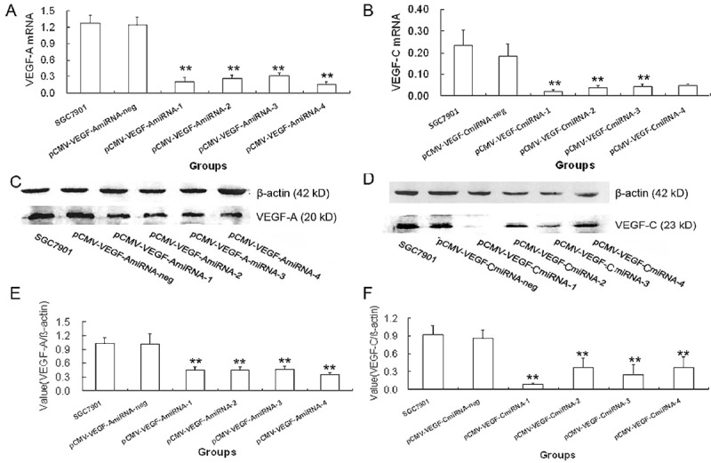
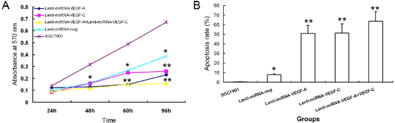
Methods: The correlations of VEGF-A and VEGF-C expressions with clinicopathologic parameters and prognosis were evaluated in patients with GC. Furthermore, lentivirus-mediated RNA interfering (RNAi) targeting VEGF-A and/or VEGF-C was employed to silence their expressions in SGC7901 GC cell line. Cell proliferation and apoptosis were measured in vitro. Suppressive effect lentivirus-mediated VEGF-A and/or VEGF-C silencing on GC growth was evaluated in GC bearing mice.

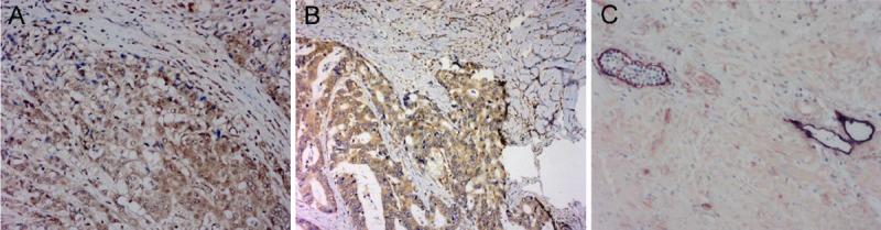
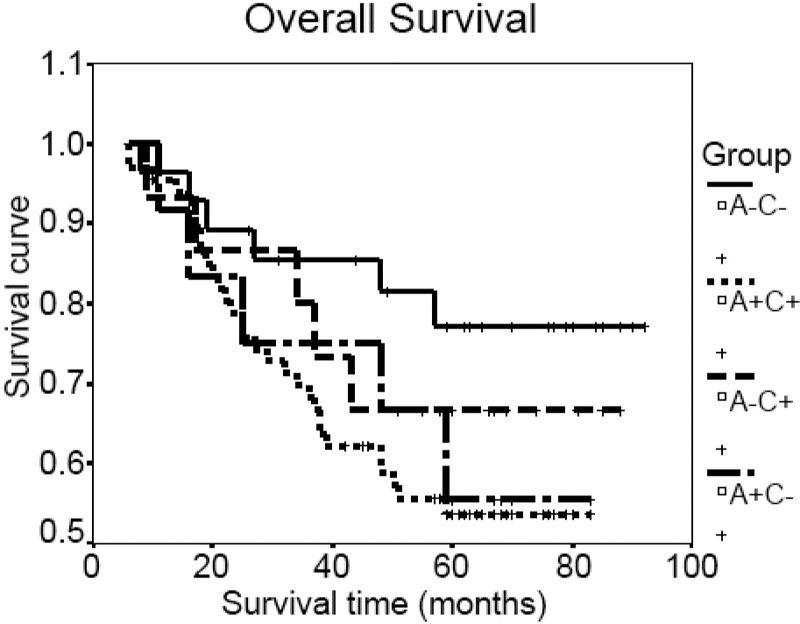
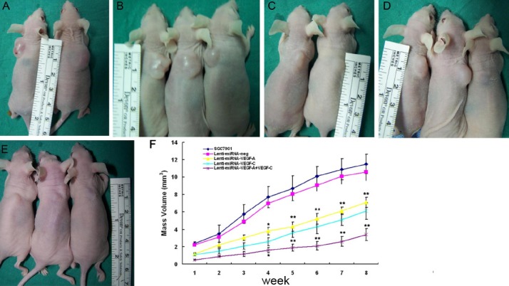
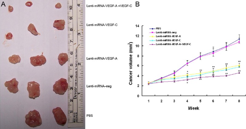
Results: The patients with high expression of both VEGF-A and VEGF-C (A+C+) had larger tumor size, higher peritumoral lymphatic vessel density(P-LVD), microvessel density(MVD), lymphatic vessel invasion (LVI), lymph node(LN) metastasis, and worse prognosis than those with low expression of both VEGF-A and VEGF-C (P<0.05). Lentivirus-mediated RNAi significantly reduced the mRNA and protein expression of VEGF-A and VEGF-C in the SGC7901 cells. The Lenti-miRNA-VEGF-A+VEGF-C significantly inhibited the cell proliferation and tumor growth, compared with Lenti-miRNA-VEGF-A or Lenti-miRNA-VEGF-C (P<0.05). In addition, Lenti-miRNA- VEGF-A+VEGF-C markedly lowered the tumor size in vivo in comparison with Lenti-miRNA-VEGF-A or Lenti-miRNA-VEGF-C (P<0.05).

Conclusion: Expressions of both VEGF-A and VEGF-C predict worse prognosis of GC patients. Combined silencing of VEGF-A and VEGF-C markedly suppresses cancer growth than silencing of VEGF-A or VEGF-C. Thus, to inhibit the expressions of VEGF-A and VEGF-C may become a novel strategy for the treatment of GC.

Keywords: Vascular endothelial growth factor-A; gastric cancer; prognosis; tumor growth; vascular endothelial growth factor-C.


Figures

Figure 1
Figure 1
Detection of expressions of VEGF-A and VEGF-C in GC by immunohistochemistry and double immunohistochemical staining for D2-40 and CD34. A. VEGF-A expression in the cytoplasm (×200). B. VEGF-C expression in the cytoplasm (×400). C. The lymphatic vessels were clearly distinguished from blood vessels after double immunohistochemical staining for D2-40 (dark purple) and CD34 (intense red) (×400).
Figure 2
Figure 2
Relationship between expressions of VEGF-A and VEGF-C and overall survival.
Figure 3
Figure 3
Expression of VEGF-A or VEGF-C in SGC7901 GC cells transduced with pCMV-VEGF-A miRNA or pCMV-VEGF-C miRNA expression plasmids at 48 h (*P<.05, **P<.01). mRNA and protein expressions of VEGF-A in SGC7901 cells transduced with pCMV-VEGF-A miRNA expression plasmids. A. Real-time qPCR showed VEGF-A mRNA expression in pCMV-VEGF-A miRNA-4 transduced cells was significantly reduced by 85.6% when compared with parental SGC7901 cells. C. Western blot assay. E. VEGF-A protein expression in pCMV-VEGF-A miRNA-4 transduced cells was significantly reduced by 89.8% when compared with parental SGC7901 cells. mRNA and protein expressions of VEGF-C in SGC7901 cells transduced with pCMV-VEGF-C miRNA expression plasmids. B. Real-time qPCR assay showed VEGF-C mRNA expression in pCMV-VEGF-C miRNA-1 transduced cells was significantly reduced by 89.8% when compared with parental SGC7901 cells. D. Western blot assay. F. VEGF-C protein expression in pCMV-VEGF-C miRNA-1 transduced cells was significantly reduced by 60.4% when compared with parental SGC7901 cells.
Figure 4
Figure 4
Combined silencing of both VEGF-A and VEGF-C significantly suppressed the cell proliferation and induced the apoptosis of GA cells. A. Cell proliferation was significantly inhibited in SGC7901 cells after infection with Lenti-miRNA-VEGF-A + Lenti-miRNA-VEGF-C vectors as measured by MTT assay, compared with control groups (*P<.05, **P<.01). B. Cells infected with Lenti-miRNA-VEGF-A + Lenti-miRNA-VEGF-C vectors had the highest apoptosis rate detected by apoptosis assay, compared with control groups (*P<.05, **P<.01).
Figure 5
Figure 5
Combined silencing of VEGF-A and VEGF-C significantly inhibited cancer growth in vivo. A. SGC7901; B. Lenti-miRNA-neg; C. Lenti-miRNA-VEGF-A; D. Lenti-miRNA- VEGF-C; E. Lenti-miRNA-VEGF-A + Lenti-miRNA-VEGF-C. F. Silencing of VEGF-A or VEGF-C in SGC7901 cells could significantly inhibit the cancer growth, when compared with the parental cells or cells infected with Lenti-miRNA-neg (*P<.05, **P<.01). The combined silencing of VEGF-A and VEGF-C could more effectively suppress the GC growth than the silencing of VEGF-A or VEGF-C alone (*P<.05, **P<.01).
Figure 6
Figure 6
Effects of Lenti-miRNA-VEGF-A and/or Lenti-miRNA-VEGF-C on cancer growth in GC bearing nude mice. A. Cancer volumes were significantly reduced in mice treated with Lenti-miRNA-VEGF-A, Lenti-miRNA-VEGF-C, or Lenti-miRNA-VEGF-A +VEGF-C, as compared to those treated with Lenti-miRNA–neg or PBS (*P<.05, **P<.01). B. Cancers in the mice treated with Lenti-miRNA-VEGF-A + VEGF-C had the smallest volumes than those treated with Lenti-miRNA-VEGF-A or Lenti-miRNA–VEGF-C (*P<.05, **P<.01).

Similar articles

Cited by

References

    1. Takahashi S. Vascular endothelial growth factor (VEGF), VEGF receptors and their inhibitors for antiangiogenic tumor therapy. Biol Pharm Bull. 2011;34:1785–1788. - PubMed
    1. Fondevila C, Metges JP, Fuster J, Grau JJ, Palacin A, Castells A, Volant A, Pera M. p53 and VEGF expression are independent predictors of tumour recurrence and survival following curative resection of gastric cancer. Br J Cancer. 2004;90:206–215. - PMC - PubMed
    1. Aoyagi K, Kouhuji K, Yano S, Miyagi M, Imaizumi T, Takeda J, Shirouzu K. VEGF significance in peritoneal recurrence from gastric cancer. Gastric Cancer. 2005;8:155–163. - PubMed
    1. Bjorndahl MA, Cao R, Burton JB, Brakenhielm E, Religa P, Galter D, Wu L, Cao Y. Vascular endothelial growth factor-a promotes peritumoral lymphangiogenesis and lymphatic metastasis. Cancer Res. 2005;65:9261–9268. - PubMed
    1. Shida A, Fujioka S, Ishibashi Y, Kobayashi K, Nimura H, Mitsumori N, Suzuki Y, Kawakami M, Urashima M, Yanaga K. Prognostic significance of vascular endothelial growth factor D in gastric carcinoma. World J Surg. 2005;29:1600–1607. - PubMed

MeSH terms

Substances