

A RTK-based functional RNAi screen reveals determinants of PTX-3 expression
- PMID: 23573312
- PMCID: PMC3606855
A RTK-based functional RNAi screen reveals determinants of PTX-3 expression
Abstract
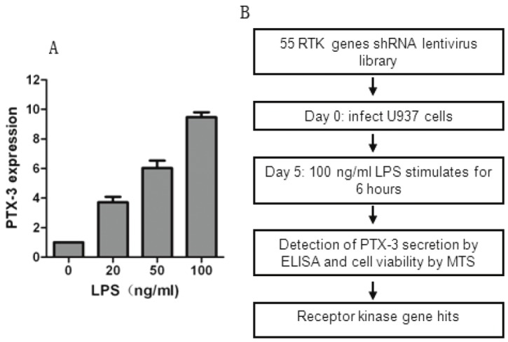
Aim: The aim of the present study was to explore the role of receptor tyrosine kinases (RTKs) in the regulation of expression of PTX-3, a protector in atherosclerosis.
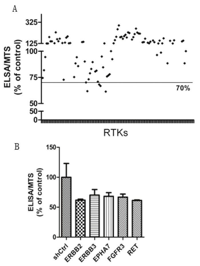
Methods: Human monocytic U937 cells were infected with a shRNA lentiviral vector library targeting human RTKs upon LPS stimuli and PTX-3 expression was determined by ELISA analysis. The involvement of downstream signaling in the regulation of PTX-3 expression was analyzed by both Western blotting and ELISA assay.
Results: We found that knocking down of ERBB2/3, EPHA7, FGFR3 and RET impaired PTX-3 expression without effects on cell growth or viability. Moreover, inhibition of AKT, the downstream effector of ERBB2/3, also reduced PTX-3 expression. Furthermore, we showed that FGFR3 inhibition by anti-cancer drugs attenuated p38 activity, in turn induced a reduction of PTX-3 expression.
Conclusion: Altogether, our study demonstrates the role of RTKs in the regulation of PTX-3 expression and uncovers a potential cardiotoxicity effect of RTK inhibitor treatments in cancer patients who have symptoms of atherosclerosis or are at the risk of atherosclerosis.
Keywords: PTX-3; RNAi screening; RTKs; atherosclerosis; cardiotoxicity; target therapy.
Figures


Similar articles
-
Pentraxin-3 Is a TSH-Inducible Protein in Human Fibrocytes and Orbital Fibroblasts.Endocrinology. 2015 Nov;156(11):4336-44. doi: 10.1210/en.2015-1399. Epub 2015 Aug 19. Endocrinology. 2015. PMID: 26287404 Free PMC article.
-
Differential expression of receptor tyrosine kinases (RTKs) and IGF-I pathway activation in human uterine leiomyomas.Mol Med. 2008 May-Jun;14(5-6):264-75. doi: 10.2119/2007-00101.Yu. Mol Med. 2008. PMID: 18231572 Free PMC article.
-
Pentraxin 3 promotes oxLDL uptake and inhibits cholesterol efflux from macrophage-derived foam cells.Exp Mol Pathol. 2014 Jun;96(3):292-9. doi: 10.1016/j.yexmp.2014.03.007. Epub 2014 Mar 24. Exp Mol Pathol. 2014. PMID: 24675235
-
Receptor Tyrosine Kinases: Translocation Partners in Hematopoietic Disorders.Trends Mol Med. 2017 Jan;23(1):59-79. doi: 10.1016/j.molmed.2016.11.002. Epub 2016 Dec 14. Trends Mol Med. 2017. PMID: 27988109 Review.
-
Proteolytic cleavage, trafficking, and functions of nuclear receptor tyrosine kinases.FEBS J. 2015 Oct;282(19):3693-721. doi: 10.1111/febs.13342. Epub 2015 Jul 4. FEBS J. 2015. PMID: 26096795 Free PMC article. Review.
Cited by
-
A key mediator, PTX3, of IKK/IκB/NF-κB exacerbates human umbilical vein endothelial cell injury and dysfunction.Int J Clin Exp Pathol. 2014 Oct 15;7(11):7699-707. eCollection 2014. Int J Clin Exp Pathol. 2014. PMID: 25550806 Free PMC article.
-
Pentraxin-3 as a prognostic marker in patients with small-cell lung cancer.Med Oncol. 2014 Oct;31(10):207. doi: 10.1007/s12032-014-0207-1. Epub 2014 Sep 2. Med Oncol. 2014. PMID: 25178940
-
Candidate SNP Markers of Atherogenesis Significantly Shifting the Affinity of TATA-Binding Protein for Human Gene Promoters show stabilizing Natural Selection as a Sum of Neutral Drift Accelerating Atherogenesis and Directional Natural Selection Slowing It.Int J Mol Sci. 2020 Feb 5;21(3):1045. doi: 10.3390/ijms21031045. Int J Mol Sci. 2020. PMID: 32033288 Free PMC article.
References
-
- Seidman A, Hudis C, Pierri MK, Shak S, Paton V, Ashby M, Murphy M, Stewart SJ, Keefe D. Cardiac dysfunction in the trastuzumab clinical trials experience. J. Clin. Oncol. 2002;20:1215–1221. - PubMed
-
- Chu TF, Rupnick MA, Kerkela R, Dallabrida SM, Zurakowski D, Nguyen L, Woulfe K, Pravda E, Cassiola F, Desai J, George S, Morgan JA, Harris DM, Ismail NS, Chen JH, Schoen FJ, Van den Abbeele AD, Demetri GD, Force T, Chen MH. Cardiotoxicity associated with tyrosine kinase inhibitor sunitinib. Lancet. 2007;370:2011–2019. - PMC - PubMed
-
- Escudier B, Eisen T, Stadler WM, Szczylik C, Oudard S, Siebels M, Negrier S, Chevreau C, Solska E, Desai AA, Rolland F, Demkow T, Hutson TE, Gore M, Freeman S, Schwartz B, Shan M, Simantov R, Bukowski RM TARGET Study Group. Sorafenib in advanced clear-cell renalcell carcinoma. N Engl J Med. 2007;356:125–134. - PubMed
-
- De Keulenaer GW, Doggen K, Lemmens K. The vulnerability of the heart as a pluricellular paracrine organ: lessons from unexpected triggers of heart failure in targeted ErbB2 anticancer therapy. Circ Res. 2010;106:35–46. - PubMed
Publication types
MeSH terms
Substances
LinkOut - more resources
Full Text Sources
Medical
Molecular Biology Databases
Research Materials
Miscellaneous