

Inflammatory angiomyolipoma of the liver: an unusual case suggesting relationship to IgG4-related pseudotumor
- PMID: 23573326
- PMCID: PMC3606869
Inflammatory angiomyolipoma of the liver: an unusual case suggesting relationship to IgG4-related pseudotumor
Abstract
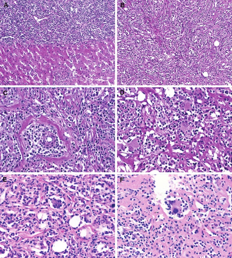
Hepatic angiomyolipoma (AML) is rare. Based on its wide histomorphological range, several distinctive histological variants have been delineated. However, hepatic AML displaying predominantly or exclusively inflammatory pattern closely mimicking inflammatory pseudotumor (IPT) is exceptionally rare with only 7 cases reported so far. We herein describe a new case of hepatic inflammatory AML in a 51-year-old woman who presented with unexplained constitutional symptoms suggesting an infectious disease. A liver mass was detected during imaging examination and resected (4.3 cm in maximum diameter). The patient's symptoms resolved completely after surgery. Currently, she is alive and well 7 years after surgery. She has no evidence of other organ manifestations of IgG4-related systemic disease. The tumor displayed a pure IPT-like histological pattern with dense infiltrates of plasma cells, lymphocytes and histiocytes admixed with scattered few adipocytes, irregularly distributed thick-walled vessels (some of them showed obliterative phlebitis) as well as aggregates and fascicles of histiocytoid and spindle-shaped myoid cells that were immunoreactive for HMB45 and Melan A with focal expression of alpha smooth muscle actin. Lesional cells were negative for desmin, protein S100, CD21, CD23, CD15, CD30, HepPar-1, pankeratin (KL-1), ALK1, and EBV in situ hybridization (EBER). The surrounding liver parenchyma showed striking lymphoplasmacytic non-destructive pericholangitis. Numerous scattered and aggregated IgG4 positive plasma cells were seen within the mass and in the peritumoral inflammatory lesions (mean, 37 cells/HPF; IgG4: IgG ratio = 28%). To our knowledge, this is the first report of hepatic inflammatory AML closely resembling IgG4-related IPT of the liver. A possible role for IgG4 seems likely to explain the peculiar histological features and the unusual clinical presentation in this case.
Keywords: Angiomyolipoma; IgG4; cholangitis; inflammatory; inflammatory pseudotumor; liver.
Figures


References
-
- Ishak KG. Mesenchymal tumors of the liver. In: Okuda K, Peters RL, editors. Hepatocellular Carcinoma. New York: John Wiley & Sons; 1976.
-
- Goodman ZD, Ishak KG. Angiomyolipomas of the liver. Am J Surg Pathol. 1984;8:745–50. - PubMed
-
- Chang Z, Zhang JM, Ying JQ, Ge YP. Characteristics and treatment strategy of hepatic angiomyolipoma: a series of 94 patients collected from four institutions. J Gastrointestin Liver Dis. 2011;20:65–9. - PubMed
-
- Nonomura A, Enomoto Y, Takeda M, Takano M, Morita K, Kasai T. Angiomyolipoma of the liver: a reappraisal of morphological features and delineation of new characteristic histological features from the clinicopathological findings of 55 tumours in 47 patients. Histopathology. 2012;61:863–80. - PubMed
Publication types
MeSH terms
Substances
LinkOut - more resources
Full Text Sources
Medical