. 2013 May 1;12(9):1433-49.
doi: 10.4161/cc.24550. Epub 2013 Apr 10.

Progesterone receptors induce FOXO1-dependent senescence in ovarian cancer cells

Affiliations

Progesterone receptors induce FOXO1-dependent senescence in ovarian cancer cells

Caroline H Diep et al. Cell Cycle. .

Abstract

Loss of nuclear progesterone receptors (PR) and low circulating progesterone levels are associated with increased ovarian cancer (OC) risk. However, PR are abundantly expressed in a significant percentage of serous and endometrioid ovarian tumors; patients with PR+ tumors typically experience longer progression-free survival relative to those with PR-null tumors. The molecular mechanisms of these protective effects are poorly understood. To study PR action in OC in the absence of added estrogen (i.e., needed to induce robust PR expression), we created ES-2 OC cells stably expressing vector control or GFP-tagged PR-B (GFP-PR). Progestin (R5020) stimulation of ES-2 cells stably expressing GFP-PR induced cellular senescence characterized by altered cellular morphology, prolonged survival, senescence-associated β-galactosidase activity, G1 cell cycle arrest and upregulation of the cell cycle inhibitor, p21, as well as the Forkhead-box transcription factor, FOXO1; these results repeated in unmodified ER+/PR+ PEO4 OC cells. PR-B and FOXO1 were detected within the same PRE-containing regions of the p21 upstream promoter. Knockdown of p21 resulted in molecular compensation via FOXO1-dependent upregulation of numerous FOXO1 target genes (p15, p16, p27) and an increased rate of senescence. Inhibition of FOXO1 (with AS1842856) or stable FOXO1 knockdown inhibited progestin-induced p21 expression and blocked progestin-induced senescence. Overall, these findings support a role for PR as a tumor suppressor in OC cells, which exhibits inhibitory effects by inducing FOXO1-dependent cellular senescence. Clinical "priming" of the PR-FOXO1-p21 signaling pathway using PR agonists may provide a useful strategy to induce irreversible cell cycle arrest and thereby sensitize OC cells to existing chemotherapies as part of combination "two-step" therapies.

Keywords: AS1842856; FOXO1; breast cancer; forkhead transcription factor; ovarian cancer; p21; progesterone receptor; progestin; senescence.


Figures
None
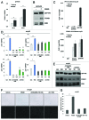
Figure 1. PR is expressed in human ovarian cancer tissues and cancer cell lines. (A) Immunohistochemical staining for PR in human ovarian cancer tissues representing the five major sub-types of ovarian surface epithelial (OSE) origin (n = 504). (B) RT-qPCR analysis of PR mRNA expression in a panel of six ovarian cancer cell lines and one immortalized, non-transformed cell line relative to PR+ T47D breast cancer cells. All values were normalized to GAPDH levels. (C) RT-qPCR analysis of PR mRNA expression of PEO4 cells treated with vehicle (ethanol) or β-estradiol (E2, 1 nM) for 24 h (n = 3, **p ≤ 0.01). (D) Western blot analysis of PR and ERα protein expression in PEO4 cells treated with vehicle, R5020 (10 nM, 1 h), E2 (1 nM, 48 h) and E2 (1 nM, 48 h) followed by R5020 (10 nM, 1 h). T47D CO total cell lysate was loaded on the same gel as a positive control for PR expression. Total ERK was used as a loading control. (E) Inset, western blot analysis of PR expression in PEO4 cells treated with vehicle and R5020 (1 and 10 μM) for 48 h. RT-qPCR analysis of SGK mRNA expression after 24 h and 96 h R5020 treatment (1 and 10 μM) in PEO4 cells (n = 3, *p ≤ 0.05, **p ≤ 0.01). All values were normalized to GAPDH levels.
None
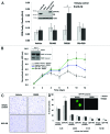
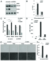
Figure 2. Stable expression of PR in ES-2 cells increases cell survival and inhibits cell colony formation. (A) Inset, western blot analysis showing stable expression of GFP-tagged PR-B in ES-2 cells (GFP-PR) as compared with parental ES-2 cells stably expressing GFP-tagged empty vector construct (empty control) and PR-B expressing T47D breast cancer cells (T47D-YB). ES-2 GFP-PR cells transiently transfected with a progesterone response element (PRE) containing luciferase reporter construct were treated for 48 h with R5020 (10 nM) or RU486 (1 uM). Relative luciferase units (RLU) were normalized to the mean result ± standard deviation (SD) for Renilla luciferase expression (n = 4, *p ≤ 0.05). (B) Inset, western blot analysis of total and cleaved PARP in GFP-PR-containing ES-2 cells treated with R5020 for 4 d. Viable GFP-PR cells continuously treated with R5020 (10 nM) as measured by MTT assay (all values normalized to day 0 readings, mean ± SD, n = 3, *p ≤ 0.05). (C) Empty control and GFP-PR expressing cells grown in soft-agar and stimulated with R5020 (10 nM) for 4 wk. Colonies were stained with crystal violet. (D) Quantification of equal numbers of colonies grown in soft-agar for 4 wk (mean ± SD, n = 3 fields/sample, 102 colonies/field, *p ≤ 0.05). Inset, representative live-colony image taken at 100× magnification demonstrating the presence of viable, single- and two-cell colonies in 4 wk R5020 (10 nM) treated GFP-PR samples.
None
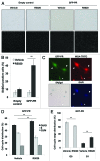
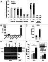
Figure 3. PR expression and activity induces cellular senescence. (A) Representative staining for SAβGal activity of empty control and GFP-PR cells treated with R5020 (10 nM) for 96 h. (magnification = 100×). Cell samples were mounted onto glass slides using ProLong® Gold Antifade Reagent with DAPI (Invitrogen) for brightfield microscopy. (B) Percentage of positive SAβGal cells was determined from quantitating three fields at 100× magnification. Values were normalized to total nuclei present in each field from DAPI staining (n = 3, **p ≤ 0.01). (C) Exposure of ES-2 cells expressing GFP-PR to R5020 (10 nM) for 12 d induced cellular senescence as indicated by cells (arrowheads) with increased SAβGal activity, while non-senescent cells (asterisk) remain SAβGal-negative. Senescent GFP-PR-containing cells also develop a characteristically larger, more flattened morphology as revealed by TRITC-labeled wheat germ agglutinin (WGA-TRITC) cell membrane staining. Nuclei were identified by DAPI counterstaining. All images were acquired at 400× magnification. (D) Cell cycle analysis by propidium iodide staining of GFP-PR-containing cells treated with R5020 (10 nM) for 96 h (n = 3, **p ≤ 0.01). (E) Flow cytometric analysis of DNA and RNA by Hoescht 33342 and Pyronin Y staining, respectively, of GFP-PR-containing cells treated with R5020 (10 nM) (**p ≤ 0.01).
None
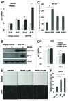
Figure 4. Progestins upregulate p21 expression to mediate cellular senescence. (A) RT-qPCR analysis of p21 mRNA levels after 24 h and 96 h R5020 (10 nM) treatment in empty control and GFP-PR-containing cells (n = 3, **p ≤ 0.01). (B) Western blot analysis of p21 protein expression in empty control and GFP-PR-containing cells after 8 d of R5020 (10 nM) treatment. (C) ES-2 GFP-PR cells transiently transfected with a p21 promoter-containing luciferase reporter construct were treated for 24 h with R5020 (10 nM) or RU486 (1 uM). Relative luciferase units (RLU) were normalized to the mean result ± standard deviation (SD) for Renilla luciferase expression (n = 4, **p ≤ 0.01). (D) RT-qPCR analysis of p21 mRNA expression after 24 h and 96 h R5020 (1 and 10 μM) treatment in PR+ PEO4 cells (n = 3, *p ≤ 0.05, **p ≤ 0.01). (E) Representative staining for SAβGal activity in PEO4 cells treated with R5020 (1 and 10 μM) for 96 h. (magnification = 100×). Cell samples were mounted onto glass slides using ProLong® Gold Antifade Reagent with DAPI (Invitrogen) for brightfield microscopy. (F) Percentage of positive SAβGal cells was determine from quantitating three fields at 100× magnification. Values were normalized to total nuclei present per field from DAPI staining (n = 3, *p ≤ 0.05).
None
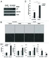
Figure 5. High p21 expression is dispensable for PR-induced cellular senescence. (A) Western blot analysis of p21 expression after lentiviral infection of shRNA oligonucleotides containing a scramble, non-targeting sequence (sh-control) or p21-targeting sequence (sh-p21). Cells were treated with R5020 (10 nM) for 8 d after stable infection of shRNA oligonucleotides. (B) Representative staining for SAβgal activity in cells expressing either sh-control or sh-p21 and treated with R5020 (10 nM) for 96 h (magnification = 100×). Cell samples were mounted onto glass slides using ProLong® Gold Antifade Reagent with DAPI (Invitrogen) for brightfield microscopy. (C) Percentage of positive Saβgal expressing cells was determined from quantitating three fields at 100× magnification. Values were normalized to total nuclei present from DAPI staining (n = 3, *p ≤ 0.05, **p ≤ 0.01). (D) RT-qPCR analysis of p15, p16, p21 and p27 mRNA expression in the sh-p21 knockdown cells treated with R5020 (10 nM) for 96 h (n = 3, *p < 0.05, **p < 0.01).
None
Figure 6. Progestin treatment of GFP-PR-containing cells stimulates FOXO1 expression and promotes PR recruitment to the p21 promoter. (A) RT-qPCR analysis of FOXO1 mRNA expression after 24 h and 96 h R5020 (10 nM) treatment of GFP-PR-containing cells (n = 3, **p ≤ 0.01). (B) Western blot analysis of FOXO1 expression in response to 96 h of R5020 (10 nM) treatment in GFP-PR-containing cells. (C) RT-qPCR analysis of PR and FOXO1 recruitment to p21. Empty control and GFP-PR expressing cells were stimulated with vehicle or R5020 (10 nM) for 1 h. Fixed lysates were chromatin immunoprecipated with antibodies to PR or FOXO1 and qRT-PCR was performed on isolated DNA. (D) RT-qPCR analysis of p15, p16, p21 and p27 mRNA expression in sh-p21 knockdown cells treated with R5020 (10 nM), FOXO1 inhibitor, AS1842856 (AS) (50 and 100 nM), or the combination of AS1842856 and R5020 for 96 h (n = 2, *p < 0.05, **p < 0.01). (E) Western blot analysis of FOXO1 expression in cells expressing sh-p21 after treatment with vehicle, R5020 (10 nM), AS1842856 (50 and 100 nM), or the combination of AS1842856 and R5020 for 96 h. (F) Representative staining for SAβGal activity of sh-p21-knockdown cells treated with R5020 (10 nM), AS1842856 (AS, 100 nM), or the combination of AS1842856 and R5020 for 96 h. (magnification = 100×). Cell samples were mounted onto glass slides using ProLong® Gold Antifade Reagent with DAPI (Invitrogen) for brightfield microscopy. (G) Percentage of positive SAβGal cells was determined from quantitating three fields at 100× magnification. Values were normalized to total nuclei present in each field from DAPI staining (n = 2, **p ≤ 0.01).
None
Figure 7. PR-induced cellular senescence is dependent on FOXO1 expression. (A) Western blot showing FOXO1 expression in cells expressing either sh-control or sh-FOXO1 after treatment of R5020 (10 nM) for 96 h. (B) RT-qPCR analysis of p21 mRNA expression in sh-control and sh-FOXO1-containing cells after 96 h of R5020 (10 nM) treatment (n = 3, **p ≤ 0.01). (C) Cell cycle analysis by propidium iodide staining of cells expressing either sh-control or sh-FOXO1 and stimulated with R5020 for 96 h. (n = 3, **p ≤ 0.01). (D) RT-qPCR analysis of PR recruitement to p21. Sh-control and sh-FOXO1-containing cells were stimulated with vehicle or R5020 (10 nM) for 1 h. Fixed lysates were chromatin immunoprecipated with an antibody to PR and qRT-PCR was performed on isolated DNA. (E) Representative staining of SAβGal activity of sh-control and sh-FOXO1-containing cells after treatment of R5020 for 96 h. Cell samples were mounted onto glass slides using ProLong® Gold Antifade Reagent with DAPI (Invitrogen) for brightfield microscopy. (F) Percentage of positive SAβGal cells were determined from quantitating three fields at 100× magnification. Values were normalized to total nuclei present from DAPI staining. (n = 3, **p ≤ 0.01).
None
Figure 8. Proposed model of PR-induced cellular senescence in ovarian cancer cells. Progestin treatment of PR-expressing OC cells induces expression of p21 and FOXO1. PR and FOXO1 cooperatively upregulate p21 expression to promote cellular senescence. In the absence of p21, PR induces numerous FOXO1 target genes that compensate as mediators of senescence.

Comment in

Similar articles

Cited by

References

    1. Atlanta: American Cancer Society Cancer Facts & Figures. 2012.
    1. Lange CA, Yee D. Progesterone and breast cancer. Womens Health (Lond Engl) 2008;4:151–62. doi: 10.2217/17455057.4.2.151. - DOI - PMC - PubMed
    1. Jacobsen BM, Richer JK, Schittone SA, Horwitz KB. New human breast cancer cells to study progesterone receptor isoform ratio effects and ligand-independent gene regulation. J Biol Chem. 2002;277:27793–800. doi: 10.1074/jbc.M202584200. - DOI - PubMed
    1. Richer JK, Jacobsen BM, Manning NG, Abel MG, Wolf DM, Horwitz KB. Differential gene regulation by the two progesterone receptor isoforms in human breast cancer cells. J Biol Chem. 2002;277:5209–18. doi: 10.1074/jbc.M110090200. - DOI - PubMed
    1. Mulac-Jericevic B, Lydon JP, DeMayo FJ, Conneely OM. Defective mammary gland morphogenesis in mice lacking the progesterone receptor B isoform. Proc Natl Acad Sci USA. 2003;100:9744–9. doi: 10.1073/pnas.1732707100. - DOI - PMC - PubMed

Publication types

MeSH terms