. 2013 May 1;12(9):1416-23.
doi: 10.4161/cc.24527. Epub 2013 Apr 9.

LXCXE-independent chromatin remodeling by Rb/E2f mediates neuronal quiescence

Affiliations

LXCXE-independent chromatin remodeling by Rb/E2f mediates neuronal quiescence

Matthew G Andrusiak et al. Cell Cycle. .

Abstract

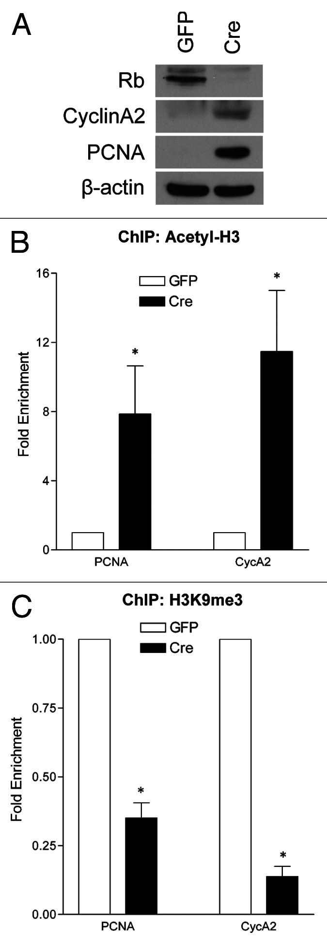
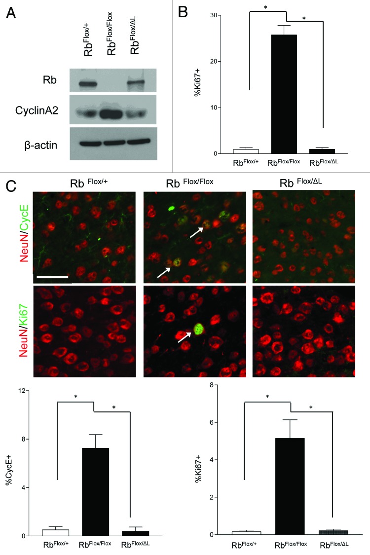
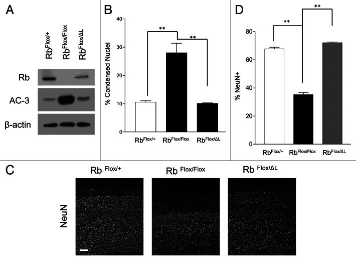
Neuronal survival is dependent upon the retinoblastoma family members, Rb1 (Rb) and Rb2 (p130). Rb is thought to regulate gene repression, in part, through direct recruitment of chromatin modifying enzymes to its conserved LXCXE binding domain. We sought to examine the mechanisms that Rb employs to mediate cell cycle gene repression in terminally differentiated cortical neurons. Here, we report that Rb loss converts chromatin at the promoters of E2f-target genes to an activated state. We established a mouse model system in which Rb-LXCXE interactions could be induciblely disabled. Surprisingly, this had no effect on survival or gene silencing in neuronal quiescence. Absence of the Rb LXCXE-binding domain in neurons is compatible with gene repression and long-term survival, unlike Rb deficiency. Finally, we are able to show that chromatin activation following Rb deletion occurs at the level of E2fs. Blocking E2f-mediated transcription downstream of Rb loss is sufficient to maintain chromatin in an inactive state. Taken together our results suggest a model whereby Rb-E2f interactions are sufficient to maintain gene repression irrespective of LXCXE-dependent chromatin remodeling.

Keywords: E2f; Rb; cell cycle; neuronal quiescence; transcription.


Figures

None
Figure 1. Acute Rb removal results chromatin activation at E2f-regulated promoters. qRT-PCR analysis of the PCNA and CycA2 promoters in GFP- or Cre-infected cortical neurons (RbFlox/Flox) at 6 DIV from chromatin immunoprecipitated DNA. (A) Western blot analysis of total protein extracted from control (GFP) and Rb-deficient (Cre) cortical neurons at 6DIV. (B) Chromatin was immunoprecipitated with an antibody directed toward acetylated histone 3 (Acetyl-H3). (C) Chromatin was immunoprecipitated using an antibody that recognizes trimethylated lysine 9 on histone 3 (H3K9me3). Error bars represent SEM (n = 3, where n represents a ChIP performed on neurons isolated from a unique embryo), significance was determined using a two-tailed Student’s t-test where *p < 0.05.
None
Figure 2. Rb regulates neuronal quiescence in an LXCXE-independent manner. (A) Western blot analysis of total protein extracted from control, Rb-deficient and RbΔL cortical neurons at 10 DIV (B) Cortical neurons were fixed at 10DIV and stained for Ki67. Percentage of Ki67+ was quantified using DAPI to represent total cell number. Error bars represent SEM (n = 3, where n represent an independent culture with multiple technical replicates); statistical significance was determined using a one-way ANOVA, where *p < 0.05 was considered statistically significant. (C) Animals of indicated genotypes were injected with tamoxifen (180 mg/kg/day i.p., 3 d) and euthanized 4 wk following the final injection. Representative pictures of ectopic expression of the cell cycle-related factors Ki67 (arrows) and Cyclin E (arrows) in neurons in the cortex of CamKCreERT2; RbFlox/Floxmice compared with CamKCreERT2; RbFlox/+ and CamKCreERT2; RbFlox/ΔLmice. Error bars represent SEM (n = 3, where n represent the cortex from a unique animal with multiple technical replicates); statistical significance was determined using a one-way ANOVA, where *p < 0.05 was considered statistically significant. Scale bar: 50 µM.
None
Figure 3. Rb regulates neuronal survival in an LXCXE independent manner. (A) Western blot analysis of total protein extracted from control, Rb-deficient and RbΔL cortical neurons at 10 DIV (B) Cortical neurons of the indicated genotypes were infected with lentiviral Cre and fixed at 10 DIV, and condensed nuclei were examined by DAPI staining. Error bars represent SEM (n = 3, where n represent an independent culture with multiple technical replicates); statistical significance was determined using a one-way ANOVA, where *p < 0.05 was considered statistically significant. (C) Animals of indicated genotypes were injected with tamoxifen (180 mg/kg/day i.p., 3 d) and euthanized 4 wk following the final injection. Representative pictures of NeuN immunofluorescence in the cortex of CamKCreERT2; RbFlox/Floxmice compared with CamKCreERT2; RbFlox/+ and CamKCreERT2; RbFlox/ΔLmice. Scale bar: 100 µm. (D) NeuN+ cells were quantified in the cortex, and percentages were obtained by normalizing to total DAPI cells for indicated genotypes. Error bars represent SEM (n = 3, where n represents the cortex from a unique animal with multiple technical replicates); statistical significance was determined using a one-way ANOVA, where *p < 0.05 was considered statistically significant.
None
Figure 4. Chromatin activation in the absence of Rb occurs at the level of E2f. qRT-PCR analysis of the PCNA and CycA2 promoters in GFP, Cre or Cre/ DP1Δ103–126 infected cortical neurons (RbFlox/Flox) at 6DIV from chromatin immunoprecipitated DNA. (A) Western blot analysis of total protein extracted from control (GFP), Rb-deficient (Cre) and Rb-deficient/dominant negative DP1 (Cre and DP1Δ103–126) cortical neurons at 6 DIV. (B) Chromatin was immunoprecipitated with an antibody directed toward acetylated histone 3 (Acetyl-H3). (C) Chromatin was immunoprecipitated using an antibody that recognizes tri-methylated lysine 9 on histone 3 (H3K9me3). Error bars represent SEM (n = 3, where n represents a ChIP performed on neurons isolated from a unique embryo); statistical significance was determined using a one-way ANOVA, where *p < 0.05 was considered statistically significant.

Comment in

References

    1. Burkhart DL, Sage J. Cellular mechanisms of tumour suppression by the retinoblastoma gene. Nat Rev Cancer. 2008;8:671–82. doi: 10.1038/nrc2399. - DOI - PMC - PubMed
    1. Dick FA. Structure-function analysis of the retinoblastoma tumor suppressor protein - is the whole a sum of its parts? Cell Div. 2007;2:26. doi: 10.1186/1747-1028-2-26. - DOI - PMC - PubMed
    1. Wang HG, Rikitake Y, Carter MC, Yaciuk P, Abraham SE, Zerler B, et al. Identification of specific adenovirus E1A N-terminal residues critical to the binding of cellular proteins and to the control of cell growth. J Virol. 1993;67:476–88. - PMC - PubMed
    1. DeCaprio JA, Ludlow JW, Figge J, Shew JY, Huang CM, Lee WH, et al. SV40 large tumor antigen forms a specific complex with the product of the retinoblastoma susceptibility gene. Cell. 1988;54:275–83. doi: 10.1016/0092-8674(88)90559-4. - DOI - PubMed
    1. Dyson N, Guida P, McCall C, Harlow E. Adenovirus E1A makes two distinct contacts with the retinoblastoma protein. J Virol. 1992;66:4606–11. - PMC - PubMed

Publication types

LinkOut - more resources