

Biosynthesis and biological function of sulfoglycolipids
- PMID: 23574804
- PMCID: PMC3669731
- DOI: 10.2183/pjab.89.129
Biosynthesis and biological function of sulfoglycolipids
Abstract
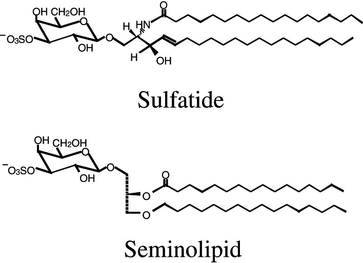
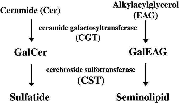
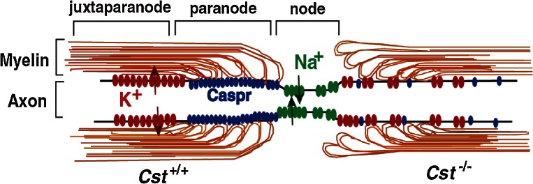
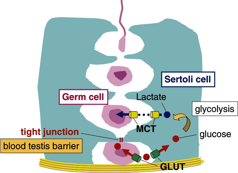
Sulfation confers negative charge on glycolipids and the attached sulfate group presents a part of determinants for the molecular interactions. Mammalian sulfoglycolipids are comprised of two major members, sulfatide (SO3-3Gal-ceramide) and seminolipid (SO3-3Gal-alkylacylglycerol). Sulfatide is abundant in the myelin sheath and seminolipid is unique to the spermatogenic cells. The carbohydrate moiety of sulfatide and seminolipid is biosynthesized via sequential reactions catalyzed by common enzymes: ceramide galactosyltransferase (CGT) and cerebroside sulfotransferase (CST). To elucidate the biological function of sulfoglycolipids, we have purified CST, cloned the CST gene, and generated CST-knockout mice. CST-null mice completely lack sulfoglycolipids all over the body. CST-null mice manifest some neurological disorders due to myelin dysfunction, an aberrant enhancement of oligodendrocyte terminal differentiation, and an arrest of spermatogenesis. CST-deficiency ameliorates L-selectin-dependent monocyte infiltration in the renal interstitial inflammation, indicating that sulfatide is an endogenous ligand of L-selectin. Studies on the molecular mechanisms underlying the biological events for which sulfoglycolipids are essential are ongoing
Figures
References
-
- Ishizuka I. (1997) Chemistry and functional distribution of sulfoglycolipids. Prog. Lipid Res. 36, 245–319 - PubMed
-
- Yamakawa T., Kiso N., Handa S., Makita A., Yokoyama S. (1962) On the structure of brain cerebroside sulfuric ester and ceramide dihexoside of erythrocytes. J. Biochem. 52, 226–227 - PubMed
-
- Ishizuka I., Suzuki M., Yamakawa T. (1973) Isolation and characterization of a novel sulfoglycolipid, ‘seminolipid,’ from boar testis and spermatozoa. J. Biochem. 73, 77–87 - PubMed
-
- Kornblatt M.J., Knapp A., Levine M., Schachter H., Murray R.K. (1974) Studies on the structure and formation during spermatogenesis of the sulfoglycerogalactolipid of rat testis. Can. J. Biochem. 52, 689–697 - PubMed
-
- Vos J.P., Lopes-Cardozo M., Gadella B.M. (1994) Metabolic and functional aspects of sulfogalactolipid. Biochim. Biophys. Acta 1211, 125–149 - PubMed
Publication types
MeSH terms
Substances
LinkOut - more resources
Full Text Sources
Other Literature Sources