Review
. 2013;89(4):139-56.
doi: 10.2183/pjab.89.139.

Synaptic inhibition and γ-aminobutyric acid in the mammalian central nervous system

Affiliations
Review

Synaptic inhibition and γ-aminobutyric acid in the mammalian central nervous system

Kunihiko Obata. Proc Jpn Acad Ser B Phys Biol Sci. 2013.

Abstract

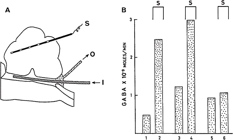
Signal transmission through synapses connecting two neurons is mediated by release of neurotransmitter from the presynaptic axon terminals and activation of its receptor at the postsynaptic neurons. γ-Aminobutyric acid (GABA), non-protein amino acid formed by decarboxylation of glutamic acid, is a principal neurotransmitter at inhibitory synapses of vertebrate and invertebrate nervous system. On one hand glutamic acid serves as a principal excitatory neurotransmitter. This article reviews GABA researches on; (1) synaptic inhibition by membrane hyperpolarization, (2) exclusive localization in inhibitory neurons, (3) release from inhibitory neurons, (4) excitatory action at developmental stage, (5) phenotype of GABA-deficient mouse produced by gene-targeting, (6) developmental adjustment of neural network and (7) neurological/psychiatric disorder. In the end, GABA functions in simple nervous system and plants, and non-amino acid neurotransmitters were supplemented.


Figures

Figure 1.
Figure 1.
Inhibitory action of GABA on mammalian CNS neuron.10) Discharges of action potentials of a Deiters neuron recorded extracellulary with a barrel of a multi-barreled electrode. Glutamic acid (anionic currents of 200 nA and 40 ms, every 1.5 s) and GABA (cationic currents of 18 nA at the time indicated) were administered iontophoretically through other barrels. GABA suppressed glutamate-induced discharges.
Figure 2.
Figure 2.
Hyperpolarization of Deiters neuron induced by GABA.34) A and C, membrane potentials recorded intracellularly from two neurons with an inner barrel of coaxial electrode. GABA or chloride ions were applied respectively with cationic or anionic currents of 500 nA through an outer barrel as indicated. Electric current artifact (B) was recorded at extracellular position and corrected for net trans-membrane potentials (dotted lines).
Figure 3.
Figure 3.
Isolated neurons for GABA assay. A, Deiters neuron attached to a glass needle in xylene.47) B, a spinal motoneuron in physiological solution.51) Parts of dendritic processes and presynaptic axons were accompanied. Scale, 50 µm.
Figure 4.
Figure 4.
GABA concentrations in single isolated Deiters neurons from non-operated (control, A and B) and operated (cerebellar vermis removed, C and D) cats.47) A and C, neurons obtained from the dorsal part of Deiters nucleus. B and D, from the ventral part. Means and S.E.M. are in parentheses. Cerebellar Purkinje axons innervate only dorsal Deiters neurons.
Figure 5.
Figure 5.
Release of GABA during stimulation of the cerebellum.49) A, arrangement for perfusion of the cat fourth ventricle. S: stimulating electrodes. I: inlet. O: outlet for the medium. B. GABA release into the fourth ventricle. Three trials in one cat. Collection period, 5 min. Cerebellar stimulation at 200 Hz were applied as indicated (S).
Figure 6.
Figure 6.
GAD67 knockout perinatal mice.66) a, wild-type (left) and KO (right) at P0.5. A KO mouse has a dilated stomach without milk (a) but lacks alveolar distension (not illustrated). b, the upper jaw. GAD67 KO mouse (right) has cleft palate (arrow, also in d). (Left) wild-type. c and d. Coronal sections of E17.5 facial region of wild-type (c) and GAD67 KO (d). n, Nasal septum. p, Palate. t, Tongue.

Similar articles

Cited by

References

    1. Sherrington, C.S. (1906) The Integrative Action of the Nervous System. Yale Univ. Press, New Haven, CT, pp. 1–413.
    1. Granit, R. (1967) Charles Scott Sherrington an Appraisal. Doubleday & Co., Garden City, NY, pp. 1–188.
    1. Eccles, J.C. (1964) The Physiology of Synapses. Springer, Berlin, pp. 1–316.
    1. Finger, S. (1994) Origins of Neuroscience. Oxford Univ. Press, New York, NY, p. 47.
    1. Setchenov, I.M. (1863) Reflexes of the Brain. (Russian pamphlet).

Substances