. 2013 Apr 10;14(4):7771-83.
doi: 10.3390/ijms14047771.

Environmental stresses induce misfolded protein aggregation in plant cells in a microtubule-dependent manner

Affiliations

Environmental stresses induce misfolded protein aggregation in plant cells in a microtubule-dependent manner

Yuko Nakajima et al. Int J Mol Sci. .

Abstract

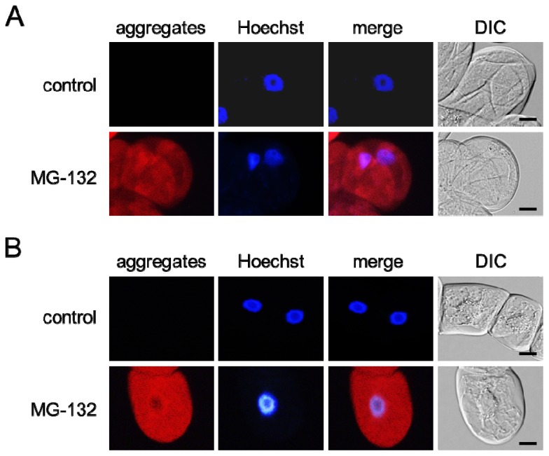
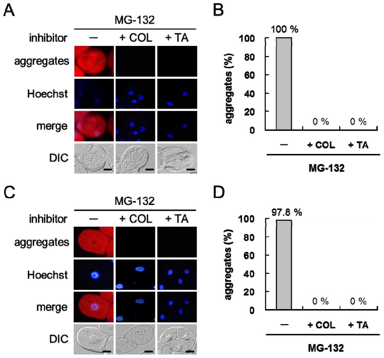
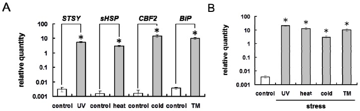
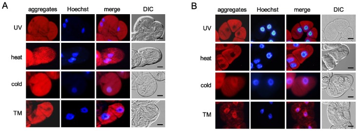
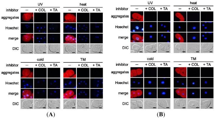
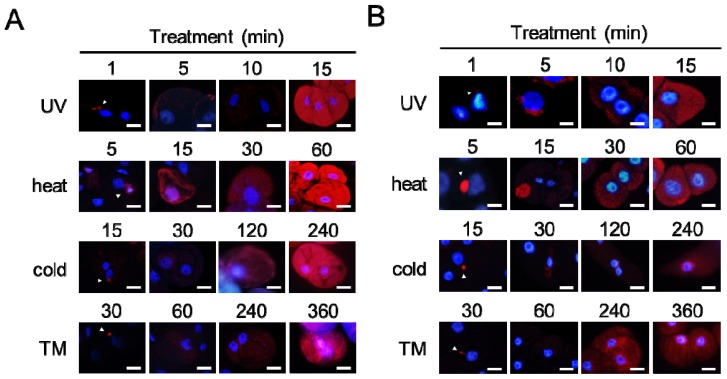
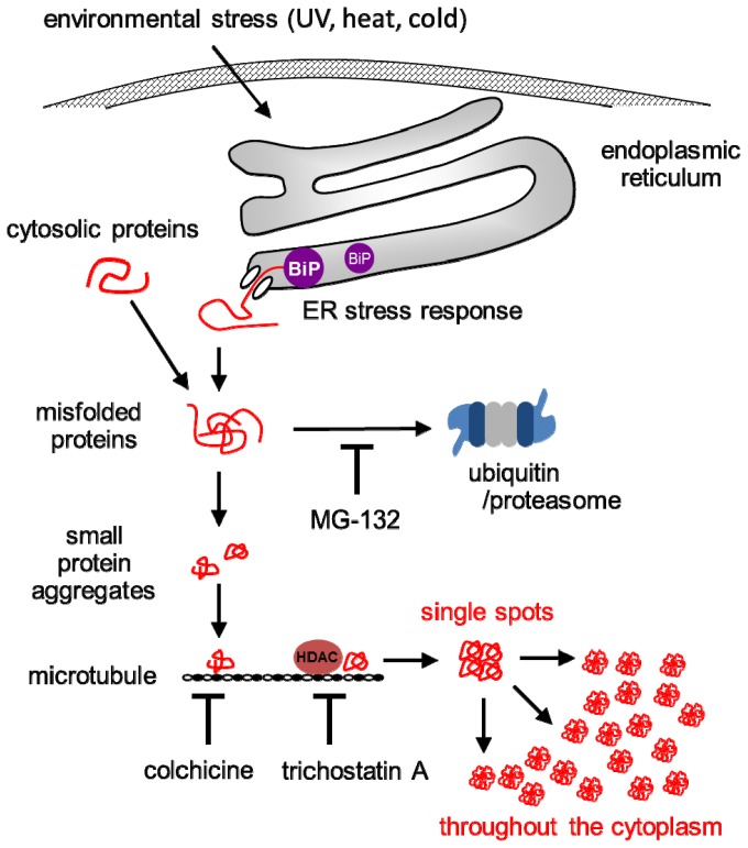
Misfolded protein aggregation in mammalian cells is one of the cellular responses to environmental stresses. However, the aggregation of misfolded proteins in plant cells exposed to environmental stresses is still poorly understood. Here, we report the misfolded protein aggregation in plant cells in response to environmental stresses, including ultraviolet (UV) radiation, heat stress and cold stress. Treatment of grape and tobacco cultured cells with MG-132, a proteasome inhibitor, induced misfolded protein aggregation. All of the environmental stresses examined induced the endoplasmic reticulum (ER) stress response in the cells. The cells under ER stress showed aggregation of misfolded proteins. The misfolded protein aggregation was completely inhibited by treatment of the cells with trichostatin A or colchicine, suggesting that the misfolded proteins might be aggregated in plant cells in a microtubule-dependent manner. Detected aggregates were initially observed immediately after exposure to the environmental stresses (1 min after UV radiation, 5 min after heat stress exposure, and 15 min after cold stress exposure). Based on these findings, we hypothesize that environmental stresses induce misfolded protein aggregation in plant cells in a microtubule-dependent manner.


Figures

Figure 1
Figure 1
Misfolded protein aggregation in plant cells. Grape (A) and tobacco (B) cultured cells were treated with MG-132. The cells were stained with Hoechst 33342 (Hoechst) and then with a ProteoStat Aggresome Detection Kit (aggregates). The red color and the blue color indicate the fluorescence of detected aggregates and stained nuclei, respectively. Merge, a merged image. DIC, an image observed under a differential-interference-contrast (DIC) microscope. Scale bars, 20 μm.
Figure 2
Figure 2
Effect of trichostatin A or colchicine treatment on misfolded protein aggregation in plant cells. (A and C) Microscopy observation; (B and D) Incidence of misfolded protein aggregation. Cotreatment with trichostatin A (TA) or colchicine (COL) and MG-132 completely suppressed misfolded protein aggregation in grape (A and B) and tobacco (C and D) cultured cells. The red color and the blue color indicate the fluorescence of detected aggregates and stained nuclei, respectively. Merge, a merged image. DIC, an image observed under a DIC microscope. Scale bars, 20 μm.
Figure 3
Figure 3
Environmental stresses induce ER stress response in plant cells. (A) Expression of stress marker genes in grape cultured cells exposed to environmental stresses. UV radiation, heat stress, and cold stress upregulated STSY, sHSP, and CBF2 expression in grape cultured cells, respectively. Tunicamycin (TM) induced luminal binding protein (BiP) expression in grape cultured cells; (B) Expression of BiP in grape cultured cells exposed to environmental stresses. All of the environmental stresses examined upregulated BiP expression in grape cultured cells similarly to TM treatment. Bars indicate means ± standard errors calculated from three independent experiments. Mean values statistically different from control (p < 0.01) are indicated by asterisks.
Figure 4
Figure 4
Environmental stresses induce misfolded protein aggregation in plant cells. (A) Grape cultured cells; (B) Tobacco cultured cells. The cells exposed to the environmental stresses were stained with Hoechst 33342 (Hoechst) and then with a ProteoStat Aggresome Detection Kit (aggregates). The red color and the blue color indicate the fluorescence of detected aggregates and stained nuclei, respectively. TM, tunicamycin; Merge, a merged image; DIC, an image observed under a DIC microscope; Scale bars, 20 μm.
Figure 5
Figure 5
Effect of trichostatin A or colchicine treatment on misfolded protein aggregation in (A) grape cultured cells and (B) tobacco cultured cells exposed to environmental stresses. Treatment with trichostatin A (TA) or colchicine (COL) completely suppressed misfolded protein aggregation induced by the environmental stresses. The red color and the blue color indicate the fluorescence of detected aggregates and stained nuclei, respectively. TM, tunicamycin; Merge, a merged image; DIC, an image observed under a DIC microscope; Scale bars, 20 μm.
Figure 6
Figure 6
Dynamics of misfolded protein aggregation in plant cells under environmental stress conditions. (A) Grape cultured cells; (B) Tobacco cultured cells. The cells were fixed at the indicated time after exposure to environmental stresses and then stained. The images are merged images. The red color and the blue color indicate the fluorescence of detected aggregates and stained nuclei, respectively. Arrowheads indicate single or small aggregates. TM, tunicamycin; Scale bars, 20 μm.
Figure 7
Figure 7
Theoretical model of misfolded protein aggregation in plant cells under environmental stress condition. Environmental stresses induce the ER stress response, including the upregulation of BiP in plant cells. Misfolded proteins are translocated from ER to the cytoplasm. Usually, misfolded proteins are degraded by proteasomes. Accordingly, misfolded protein aggregates are formed in plant cells following MG-132 treatment. Numerous misfolded proteins are likely generated in response to environmental stresses and the capacity of proteasomes to degrade them is exceeded. The aggregation of misfolded proteins occurs first in single spots and then throughout the cytoplasm. These aggregates might be formed in a microtubule-dependent manner, since colchicine and trichostatin A inhibits the aggregation. Histone deacetylase (HDAC) coordinates the formation of the aggregates on the microtubule through a yet unexplained manner(s). End lines indicate negative regulations.

References

    1. Vembar S.S., Brodsky J.L. One step at a time: Endoplasmic reticulum-associated degradation. Nature Rev. Mol. Cell Biol. 2008;9:944–957. - PMC - PubMed
    1. Kamauchi S., Nakatani H., Nakano C., Urade R. Gene expression in response to endoplasmic reticulum stress in Arabidopsis thaliana. FEBS J. 2005;272:3461–3476. - PubMed
    1. Vidair A., Huang R.N., Doxsey S.J. Heat shock causes protein aggregation and reduced protein solubility at the centrosome and other cytoplasmic locations. Int. J. Hyperth. 1996;12:12681–12695. - PubMed
    1. Buchner J. Supervising the fold: Functional principles of molecular chaperones. FASEB J. 1996;10:10–19. - PubMed
    1. Levitskaya J., Sharipo A., Leonchiks A., Ciechanover A., Masucci M.G. Inhibition of ubiquitin/proteasome-dependent protein degradation by the Gly-Ala repeat domain of the Epstein-Barr virus nuclear antigen 1. Proc. Natl. Acad. Sci. USA. 1997;94:12616–12621. - PMC - PubMed

MeSH terms

LinkOut - more resources