Advances in bladder cancer imaging
- PMID: 23574966
- PMCID: PMC3635890
- DOI: 10.1186/1741-7015-11-104
Advances in bladder cancer imaging
Abstract
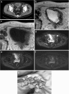
The purpose of this article is to review the imaging techniques that have changed and are anticipated to change bladder cancer evaluation. The use of multidetector 64-slice computed tomography (CT) and magnetic resonance imaging (MRI) remain standard staging modalities. The development of functional imaging such as dynamic contrast-enhanced MRI, diffusion-weighted MRI and positron emission tomography (PET)-CT allows characterization of tumor physiology and potential genotypic activity, to help stratify and inform future patient management. They open up the possibility of tumor mapping and individualized treatment solutions, permitting early identification of response and allowing timely change in treatment. Further validation of these methods is required however, and at present they are used in conjunction with, rather than as an alternative to, conventional imaging techniques.
Figures


Similar articles
-
Recent advances in imaging cancer of the kidney and urinary tract.Surg Oncol Clin N Am. 2014 Oct;23(4):863-910. doi: 10.1016/j.soc.2014.06.001. Epub 2014 Aug 13. Surg Oncol Clin N Am. 2014. PMID: 25246053 Review.
-
Bladder cancer imaging: an update.Curr Opin Urol. 2011 Sep;21(5):393-7. doi: 10.1097/MOU.0b013e32834956c3. Curr Opin Urol. 2011. PMID: 21814052 Review.
-
Genitourinary imaging: current and emerging applications.J Postgrad Med. 2010 Apr-Jun;56(2):131-9. doi: 10.4103/0022-3859.65291. J Postgrad Med. 2010. PMID: 20622393 Review.
-
Urinary bladder masses: techniques, imaging spectrum, and staging.J Comput Assist Tomogr. 2011 Jul-Aug;35(4):411-24. doi: 10.1097/RCT.0b013e31821c2e9d. J Comput Assist Tomogr. 2011. PMID: 21765295 Review.
-
[Functional imaging in bladder cancer].Urologe A. 2013 Apr;52(4):509-14. doi: 10.1007/s00120-012-3097-x. Urologe A. 2013. PMID: 23483270 German.
Cited by
-
[Oncological diseases and postoperative alterations of the bladder and urinary tract].Radiologe. 2014 Dec;54(12):1221-34; quiz 1235-6. doi: 10.1007/s00117-014-2768-6. Radiologe. 2014. PMID: 25425104 German.
-
Clinical experience of MRI in two dogs with muscle-invasive transitional cell carcinoma of the urinary bladder.J Vet Med Sci. 2016 Sep 1;78(8):1351-4. doi: 10.1292/jvms.15-0622. Epub 2016 Apr 28. J Vet Med Sci. 2016. PMID: 27149892 Free PMC article.
-
Magnetic resonance radiographic features which might lead to misdiagnosis of muscle-invasive bladder cancer based on vesical imaging reporting and data system: the application experience of a single center.Quant Imaging Med Surg. 2023 Oct 1;13(10):7258-7268. doi: 10.21037/qims-23-356. Epub 2023 Sep 11. Quant Imaging Med Surg. 2023. PMID: 37869292 Free PMC article.
-
Preoperative imaging for staging bladder cancer.Curr Urol Rep. 2015 Apr;16(4):22. doi: 10.1007/s11934-015-0496-8. Curr Urol Rep. 2015. PMID: 25724433 Review.
-
Role of Diffusion-Weighted Magnetic Resonance Imaging (DWMRI) in Assessment of Primary Penile Tumor Characteristics and Its Correlations With Inguinal Lymph Node Metastasis: A Prospective Study.World J Oncol. 2018 Nov;9(5-6):145-150. doi: 10.14740/wjon1138w. Epub 2018 Nov 23. World J Oncol. 2018. PMID: 30524639 Free PMC article.
References
-
- UK Office for National Statistics. Mortality statistics EaW. [ http://www.ons.gov.uk/ons/rel/cancer-unit/cancer-incidence-and-mortality...]
-
- Edge SB, Byrd DR, Compton CC, Fritz AG, Greene FL, Trotti A. (Eds): AJCC Cancer Staging Maual. 7. New York, NY: Springer; 2010.
-
- Jewett HJ, Strong GH. Infiltrating carcinoma of the bladder; relation of depth of penetration of the bladder wall to incidence of local extension and metastases. J Urol. 1946;55:366–372. - PubMed
-
- MacVicar AD. Bladder cancer staging. BJU Int. 2000;86(Suppl 1):111–122. - PubMed
Publication types
MeSH terms
Grants and funding
LinkOut - more resources
Full Text Sources
Other Literature Sources
Medical

