Review
. 2013 Apr 10;11(4):1203-20.
doi: 10.3390/md11041203.

A therapeutic potential for marine skeletal proteins in bone regeneration

Affiliations
Review

A therapeutic potential for marine skeletal proteins in bone regeneration

David W Green et al. Mar Drugs. .

Abstract

A vital ingredient for engineering bone tissue, in the culture dish, is the use of recombinant matrix and growth proteins to help accelerate the growth of cultivated tissues into clinically acceptable quantities. The skeletal organic matrices of calcifying marine invertebrates are an untouched potential source of such growth inducing proteins. They have the advantage of being ready-made and retain the native state of the original protein. Striking evidence shows that skeleton building bone morphogenic protein-2/4 (BMP) and transforming growth factor beta (TGF-β) exist within various marine invertebrates such as, corals. Best practice mariculture and the latest innovations in long-term marine invertebrate cell cultivation can be implemented to ensure that these proteins are produced sustainably and supplied continuously. This also guarantees that coral reef habitats are not damaged during the collection of specimens. Potential proteins for bone repair, either extracted from the skeleton or derived from cultivated tissues, can be identified, evaluated and retrieved using chromatography, cell assays and proteomic methods. Due to the current evidence for bone matrix protein analogues in marine invertebrates, together with the methods established for their production and retrieval there is a genuine prospect that they can be used to regenerate living bone for potential clinical use.
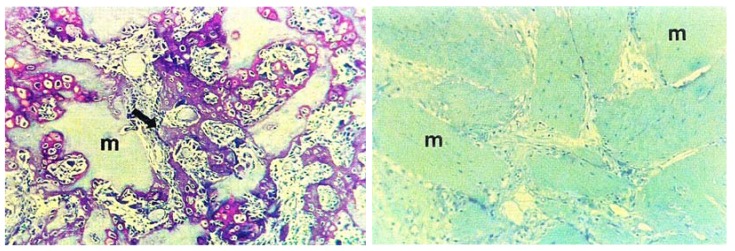
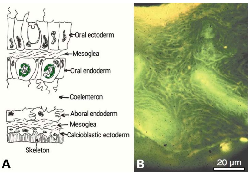

Figures

Figure 1
Figure 1
Bone formation in mammals is made possible with a bone inductive protein analogue derived from an invertebrate. In this example, a protein from Drosophila development and an analogue of skeleton building bone morphogenic protein-2/4 (BMP), was injected into the muscle of a mouse, generating new bone. The histological section on the left shows osteoinduction in living mouse subcutaneous tissue following treatment with Drosophila Decapentaplegic (dpp). New bone tissue has been reproduced shown in purple. On the right, for comparison is a histological section of untreated mouse subcutaneous tissue. (Reproduced with permission from PNAS, Sampath et al. 1993 [13]).
Figure 2
Figure 2
(A) A cross-sectional diagram through the living tissue portion of a coral and showing the interface with the exoskeleton. The calcioblastic epithelium secretes the organic matrix probably embedded within intracellular vesicles into micron or nanometric sized spaces. Biological control of mineralization is strongly implicated in corals in other ways. There are semi-permeable tight septate junctions between the cells that control ion transport and other molecules according to size and charge (After Allemand et al. [22]). The calcioblastic epithelium effectively facilitates the laying down of the inorganic skeleton; (B) Acridine orange staining of the organic matrix of Acropora sp. skeleton, which appears strong yellow at the growing region and green deeper inside the skeleton. The pale yellow regions are the centres of calcification. This microscope image was taken under polarized light and shows the global distribution of intra-skeletal organic matrices throughout the entire skeleton [25]. The coral tissue has been removed to view this organic matrix. (Reproduced with permission from the Institute of Paleobiology, Polish Academy of Sciences, Gautret et al. 2000 [25]).

References

    1. Dimitriou R., Jones E., McGonagle D., Giannoudis P.V. Bone regeneration: Current concepts and future directions. BMC Med. 2011;9:66. doi: 10.1186/1741-7015-9-66. - DOI - PMC - PubMed
    1. Romano N.H., Sengupta D., Chung C., Heilshorn S.C. Protein-engineered biomaterials: Nanoscale mimics of the extracellular matrix. Biochim. Biophys. Acta. 2011;1810:339–349. doi: 10.1016/j.bbagen.2010.07.005. - DOI - PMC - PubMed
    1. Lee K., Silva E.A., Mooney D.J. Growth factor delivery-based tissue engineering: General approaches and a review of recent developments. J. R. Soc. Interface. 2011;8:153–170. doi: 10.1098/rsif.2010.0223. - DOI - PMC - PubMed
    1. Lutolf M.P., Hubbell J.A. Synthetic biomaterials as instructive extracellular microenvironments for morphogenesis in tissue engineering. Nat. Biotechnol. 2005;23:47–55. doi: 10.1038/nbt1055. - DOI - PubMed
    1. Gillette B.M., Jensen J.A., Tang B., Yang G.J., Bazargan-Lari A., Zhong M., Sia S.K. In situ collagen assembly for integrating microfabricated three-dimensional cell-seeded matrices. Nat. Mater. 2008;7:636–640. doi: 10.1038/nmat2203. - DOI - PubMed

Substances

LinkOut - more resources