. 2013 Jun 20;88(6):154.
doi: 10.1095/biolreprod.112.106195. Print 2013 Jun.

Histopathologies, immunolocalization, and a glycan binding screen provide insights into Plasmodium falciparum interactions with the human placenta

Affiliations

Histopathologies, immunolocalization, and a glycan binding screen provide insights into Plasmodium falciparum interactions with the human placenta

Bethann S Hromatka et al. Biol Reprod. .

Abstract

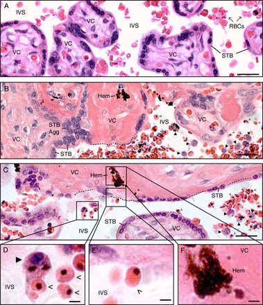
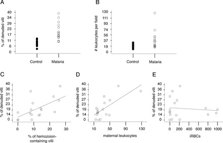
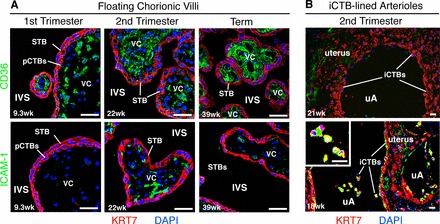
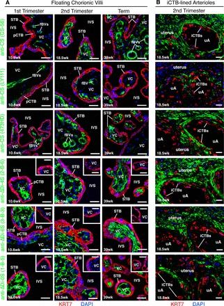
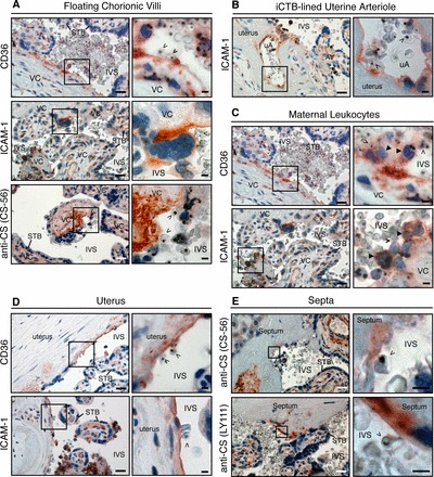
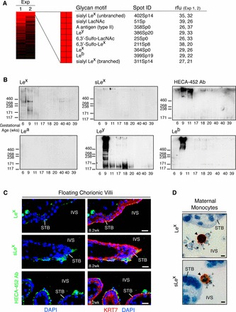
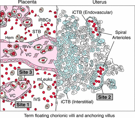
During pregnancy, Plasmodium falciparum-infected erythrocytes cytoadhere to the placenta. Infection is likely initiated at two sites where placental trophoblasts contact maternal blood: 1) via syncytiotrophoblast (STB), a multicellular transporting and biosynthetic layer that forms the surface of chorionic villi and lines the intervillous space, and 2) through invasive cytotrophoblasts, which line uterine vessels that divert blood to the placenta. Here, we investigated mechanisms of infected erythrocyte sequestration in relationship to the microanatomy of the maternal-fetal interface. Histological analyses revealed STB denudation in placental malaria, which brought the stromal cores of villi in direct contact with maternal blood. STB denudation was associated with hemozoin deposition (P = 0.01) and leukocyte infiltration (P = 0.001) and appeared to be a feature of chronic placental malaria. Immunolocalization of infected red blood cell receptors (CD36, ICAM1/CD54, and chondroitin sulfate A) in placentas from uncomplicated pregnancies showed that STB did not stain, while the underlying villous stroma was immunopositive. Invasive cytotrophoblasts expressed ICAM1. In malaria, STB denudation exposed CD36 and chondroitin sulfate A in the villous cores to maternal blood, and STB expressed ICAM1. Finally, we investigated infected erythrocyte adherence to novel receptors by screening an array of 377 glycans. Infected erythrocytes bound Lewis antigens that immunolocalized to STB. Our results suggest that P. falciparum interactions with STB-associated Lewis antigens could initiate placental malaria. Subsequent pathologies, which expose CD36, ICAM1, and chondroitin sulfate A, might propagate the infection.

Keywords: placenta; pregnancy; stroma; syncytiotrophoblast; trophoblast.


Figures

FIG. 1
FIG. 1
STB loss is a prominent histopathological feature of placental malaria. A) In control, uninfected placentas, the villous core (VC), which contains stromal cells and fetal blood vessels, is covered by a continuous layer of STB. Maternal blood cells, primarily RBCs, occupy the intervillous space (IVS). B, C) In P. falciparum-infected placentas, STB denudation (dotted lines) brought the VC into direct contact with the IVS. Some denuded villi were acellular and eosinophilic (intravillous fibrinoid; B). Some exposed VCs resembled normal stroma (C). STB aggregation (STB Agg) was commonly observed. Villi that displayed these pathological alterations often contained hemozoin (Hem). These regions were reservoirs for iRBCs (open arrowheads) and maternal leukocytes (closed arrowheads). D) Cells with the morphological appearance of monocytes (closed arrowhead) contained hemozoin and were found in close association with iRBCs (open arrowheads). Infected RBCs (E) were observed adjacent to the VCs of denuded villi (F), which often contained hemozoin. Tissue sections were stained with H&E. Bars = 50 μm (AC) and 10 μm (DF).
FIG. 2
FIG. 2
STB denudation was associated with hemozoin and maternal leukocytes. A) STB denudation (# of denuded villi per total villi) was approximately two-fold higher in the cases versus controls (SEM = 2.2%, P < 0.0001). The mean percentage of STB loss was 9.0% in controls (closed circles; range 3.0%–13.0%) and 17.0% in the malaria group (open circles; range 10.0% to 39.0%). B) Maternal leukocyte infiltration was 18% higher in the cases versus controls (SEM = 7.0%, P = 0.01). The mean number of maternal leukocytes per 400× field was 18.0 in the controls (closed circles; range 8.6–30.5) and 26.3 in the malaria group (open circles; range 14.6–128.0). CE) Linear regression analysis was used to determine associations between STB denudation and infection or inflammation. STB loss was associated with hemozoin (C; P = 0.01) and maternal leukocytes (D; P = 0.001), but not with iRBCs (E; P = 0.88).
FIG. 3
FIG. 3
CD36 and ICAM1 expression in relationship to the intervillous space during normal pregnancy. Tissue sections of placental biopsies from uncomplicated pregnancies (first-trimester, second-trimester, and term), corresponding to site 1 (A) and site 2 (B) in Supplemental Figure S1D, were analyzed. A) In floating chorionic villi at all gestational ages, anti-CD36 Ab (green, top panels) reacted with the stromal villous cores (VCs); pCTBs and STB failed to stain. ICAM1 expression (green, bottom panels), which was more variable, was not detected in first-trimester samples. Patchy staining of the VCs was evident beginning in the second trimester. No staining of the trophoblast layers was observed. B) Invasive CTB invasion of uterine arterioles (uAs) peaks in the second trimester. Therefore, these analyses focused on three placental samples that were collected from this time period. In basal plate biopsies that contained iCTB-remodeled uAs, anti-CD36 Ab (top panel) and anti-ICAM1 Ab (bottom panel) reacted with the uterine extracellular matrix (green). Invasive CTBs that lined the uAs failed to express CD36 (top panel), but stained brightly for ICAM1 (bottom panel), enlarged in inset. Biopsies were fixed in paraformaldehyde and frozen. STB and pCTBs were visualized by staining for cytokeratin 7 (KRT7, red). Nuclei were labeled with DAPI (blue). Bar = 40 μm.
FIG. 4
FIG. 4
CS-A expression is sequestered from the intervillous space during normal pregnancy. Biopsies from two placental sites (Supplemental Fig. S1D) were analyzed using the experimental strategy described in Figure 3. A) In floating chorionic villi, mAbs that specifically recognized CS-A (CS-56, LY111, 473HD) reacted with the villous cores (VCs) (green). Often, intense immunostaining was observed in association with fetal blood vessels (fBVs). The stub mAbs (2-B-6, 3-B-3, 1-B-5), which recognized epitopes exposed by chondroitinase ABC digestion, also reacted with the VCs (green). Control tissue sections that were not digested with chondroitinase ABC did not stain (insets). None of the mAbs stained STB or pCTBs, labeled with an anti-KRT7 mAb (red). B) In second-trimester basal plate biopsies that contained iCTB-remodeled uterine arterioles (uAs), the anti-CS-A mAb panel reacted with the uterine extracellular matrix, but failed to stain iCTBs that lined the uAs. Bar = 40 μm.
FIG. 5
FIG. 5
CD36, ICAM1, and CS-A expression in relationship to the intervillous space during malaria infection. A) In floating chorionic villi, villous core (VC) regions that were denuded of STB expressed CD36. STB exhibited patchy ICAM1 immunoreactivity. Abs that recognized CS-A (example shown for CS56) reacted with denuded regions (inset) and intact stroma. B) As shown for normal samples collected in San Francisco (Fig. 3B), iCTBs that lined uterine arterioles (uAs) expressed ICAM1. Anchoring villus, AV. C) Maternal leukocytes in the intervillous space (IVS) with the morphological appearance of monocytes (closed arrowheads) stained for CD36 (top panel) and ICAM1 (bottom panel). Often, these cells formed a bridge between iRBCs (open arrowheads) and STB. D) The portion of the uterus that lines the intervillous space expressed CD36 (top panel) and ICAM1 (bottom panel). E) Placental septa (Supplemental Fig. S1B) that were continuous with the intervillous space stained for CS-A (detected with CS-56 and LY111 mAbs). The brown color represents the binding of primary Abs. Sections were counterstained with hematoxylin (blue). Boxed area in left panel is enlarged as right panel. Bars = 50 μm for lower magnification and 10 μm for higher magnification micrographs.
FIG. 6
FIG. 6
A glycan binding screen suggests a role for Le carbohydrate antigens in cytoadhesion. A) Fluorescently labeled iRBCs were bound to a glycan array. An annotated heat map of potential P. falciparum binding partners that were detected in both experiments (Exp 1 2) is shown. These included a preponderance of Le blood group structures. Relative fluorescent units (rfu) were computed as the foreground minus background intensity. B) STB microvillous membranes were isolated from placentas of the gestational ages shown, separated by SDS-PAGE, and probed with mAbs that recognized the glycans that iRBCs bound on the arrays. The apparent molecular masses of markers (shown as Mr × 10−3) are on the left. First-trimester placentas had appreciable levels of Lex, sLex, and sulfated Lex (detected with HECA-452 Ab) expression (top blots). Ab reactivity declined with advancing gestational age. In contrast, Lea expression was not detected (bottom, left). Ley (bottom, middle) immunoreactivity was observed in most samples collected during the first half of pregnancy, while Leb (bottom, right) expression was primarily confined to a 9-wk preparation. C) First-trimester placental biopsies from uncomplicated pregnancies were fixed in paraformaldehyde and frozen. In floating chorionic villi, STB exhibited patchy Lex (top panel) and sLex (middle panel) expression (green). STB uniformly stained for sulfated sLex (green, bottom panel). Bar = 20 μm. D) P. falciparum-infected, term placentas were formalin fixed and paraffin embedded. Maternal leukocytes with the morphological appearance of monocytes (closed arrowheads) expressed Lex (top panel) and sLex (bottom panel). Infected RBCs (open arrowheads) were found in close association with these cells. Bar = 40 μm.
FIG. 7
FIG. 7
A model for P. falciparum sequestration by the human placenta. At a microanatomical level, P. falciparum infection changes the repertoire of placental tissues that are in direct contact with maternal blood. In this context, binding to STB (site 1) and/or iCTBs that line uterine vessels (site 2) initiates sequestration. At later stages, STB denudation brings site 3, the villous core (VC), into direct contact with maternal blood. At a molecular level, sequestration might be initiated when iRBCs adhere to STB via currently unidentified molecules and/or to endovascular iCTBs that line uterine arterioles and express ICAM1 (site 2). As infection progresses, cytoadherence to STB might also occur via ICAM1. Advanced stages of placental malaria are associated with hemozoin (Hem) and STB denudation, which exposes villous CD36 and CS-A to maternal blood (site 3). Placenta-associated maternal leukocytes (mLeuk) could also participate in adherence because they express CD36, ICAM1, and Le antigens.

References

    1. Desai M, ter Kuile FO, Nosten F, McGready R, Asamoa K, Brabin B, Newman RD. Epidemiology and burden of malaria in pregnancy. Lancet Infect Dis 2007; 7: 93 104. - PubMed
    1. Rogerson SJ, Mwapasa V, Meshnick SR. Malaria in pregnancy: linking immunity and pathogenesis to prevention. Am J Trop Med Hyg 2007; 77: 14 22. - PubMed
    1. Rogerson SJ, Hviid L, Duffy PE, Leke RF, Taylor DW. Malaria in pregnancy: pathogenesis and immunity. Lancet Infect Dis 2007; 7: 105 117. - PubMed
    1. Brabin BJ, Romagosa C, Abdelgalil S, Menendez C, Verhoeff FH, McGready R, Fletcher KA, Owens S, D'Alessandro U, Nosten F, Fischer PR, Ordi J. The sick placenta—the role of malaria. Placenta 2004; 25: 359 378. - PubMed
    1. Brabin B, Johnson P. Placental malaria and pre-eclampsia through the looking glass backwards? J Reprod Immunol 2005; 65: 1 15. - PubMed

Publication types

MeSH terms