Review
. 2013 Apr 10:11:11.
doi: 10.1186/1477-3155-11-11.

Microtechnologies to fuel neurobiological research with nanometer precision

Affiliations
Review

Microtechnologies to fuel neurobiological research with nanometer precision

Cecilia A Brunello et al. J Nanobiotechnology. .

Abstract

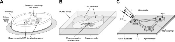
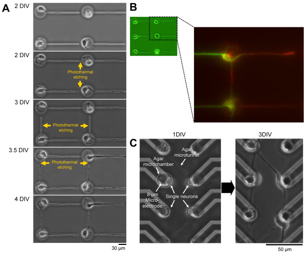
The interface between engineering and molecular life sciences has been fertile ground for advancing our understanding of complex biological systems. Engineered microstructures offer a diverse toolbox for cellular and molecular biologists to direct the placement of cells and small organisms, and to recreate biological functions in vitro: cells can be positioned and connected in a designed fashion, and connectivity and community effects of cells studied. Because of the highly polar morphology and finely compartmentalized functions of neurons, microfabricated cell culture systems and related on-chip technologies have become an important enabling platform for studying development, function and degeneration of the nervous system at the molecular and cellular level. Here we review some of the compartmentalization techniques developed so far to highlight how high-precision control of neuronal connectivity allows new approaches for studying axonal and synaptic biology.


Figures

Figure 1
Figure 1
Three techniques for isolation of axons in neuronal cell culture. (A) The Campenot chamber is a Teflon ring placed on a layer of silicon grease on top of regular cell culture dish or glass coverslip. Dissociated peripheral neurons such as dorsal root ganglion cells are placed in the middle reservoir. NGF placed in the axonal chambers attracts axons to grow through the silicon grease layer while the cell somas are restricted to the middle reservoir. (B) Microfluidic devices are typically microfabricated from PDMS. The two cell reservoirs are connected via microgrooves (typically appr. 3–10 μm width and hight, >450 μm in length). Axons grow through the microgrooves while somas and dendrites are limited in the cell reservoirs. Many types of neurons including central neurons can be grown in microfluidic devices. (C) Individual neurons can be cultured to create directed neuronal networks using the low-melting agar etching technique. Glass coverslip is coated with a 50 nm-thick indium-tin iodide (ITO) layer below the agar. ITO allows highly localized photo-thermal etching of agar with a 1064-nm infrared laser beam. Based on observed initial axon growth, microchannels can be opened to connect individual neurons to form simple networks. The panel C is reproduced from [7].
Figure 2
Figure 2
Topographic control of axon/dendrite connections by stepwise formation of neuronal network pattern using photothermal etching. By applying stepwise photo-thermal etching to agar microchambers during cultivation, the direction of synaptic connectivity in a living neuronal network can be controlled. This allows the tunnels in which axons were elongated to be flexibly extended by melting the narrow micrometer-order grooves (microchannels) in steps through photo-thermal etching where a portion of the agar layer is melted with infrared laser beam. (A) Phase-contrast images of single hippocampal neurons during the stepwise etching procedure. When cultivation started, single cells were placed into the agar microchambers and cultivated for 2 days in vitro (DIV). The first single neurites that elongate from the cells into the microchannels are typically axons, whereas the second and latter neurites are typically dendrites. When the elongations of neurites are sufficiently stable, additional photo-thermal etching steps can be applied to connect two adjacent agar microchambers. (B) Fluorescent staining of neurites with axon- and dendrite-specific markers at DIV 5 (red, synapsin I for axons; green, MAP2 for dendrites). (C) A combination of microelectrode and agar microchambers for an individual-cell-based neural network patterning and electrophysiological recording. 8-μm microelectrode-size agar microchambers and 2-μm microtunnels were arranged on the MEA chip to control the positions of neurons and their connections.

References

    1. Hammarback JA, Palm SL, Furcht LT, Letourneau PC. Guidance of neurite outgrowth by pathways of substratum-adsorbed laminin. J Neurosci Res. 1985;13:213–220. doi: 10.1002/jnr.490130115. - DOI - PubMed
    1. Wu HI, Cheng GH, Wong YY, Lin CM, Fang W, Chow WY, Chang YC. A lab-on-a-chip platform for studying the subcellular functional proteome of neuronal axons. Lab Chip. 2010;10:647–653. doi: 10.1039/b918217a. - DOI - PubMed
    1. Campenot RB. Local control of neurite development by nerve growth factor. Proc Natl Acad Sci U S A. 1977;74:4516–4519. doi: 10.1073/pnas.74.10.4516. - DOI - PMC - PubMed
    1. McDonald JC, Whitesides GM. Poly(dimethylsiloxane) as a material for fabricating microfluidic devices. Acc Chem Res. 2002;35:491–499. doi: 10.1021/ar010110q. - DOI - PubMed
    1. Taylor AM, Blurton-Jones M, Rhee SW, Cribbs DH, Cotman CW, Jeon NL. A microfluidic culture platform for CNS axonal injury, regeneration and transport. Nat Methods. 2005;2:599–605. doi: 10.1038/nmeth777. - DOI - PMC - PubMed

Publication types

LinkOut - more resources