. 2013 May 16;497(7449):332-7.
doi: 10.1038/nature12107. Epub 2013 Apr 10.

Structural and molecular interrogation of intact biological systems

Affiliations

Structural and molecular interrogation of intact biological systems

Kwanghun Chung et al. Nature. .

Abstract

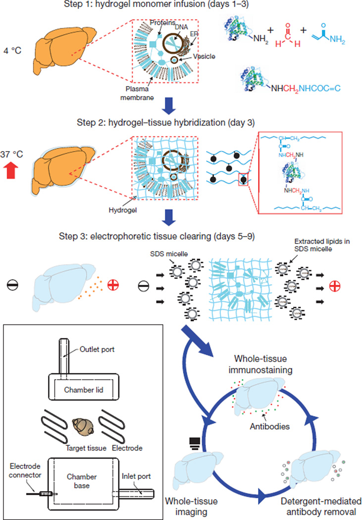
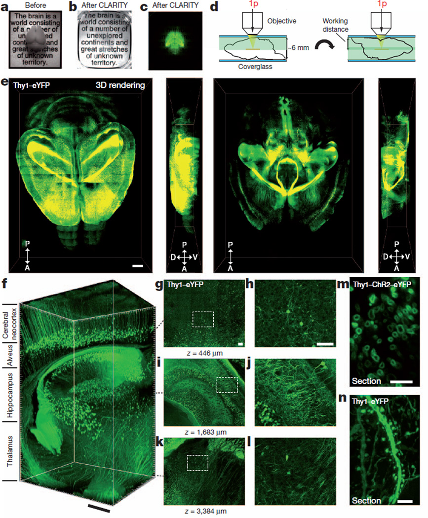
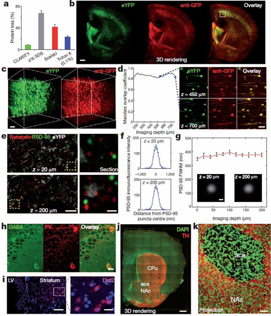
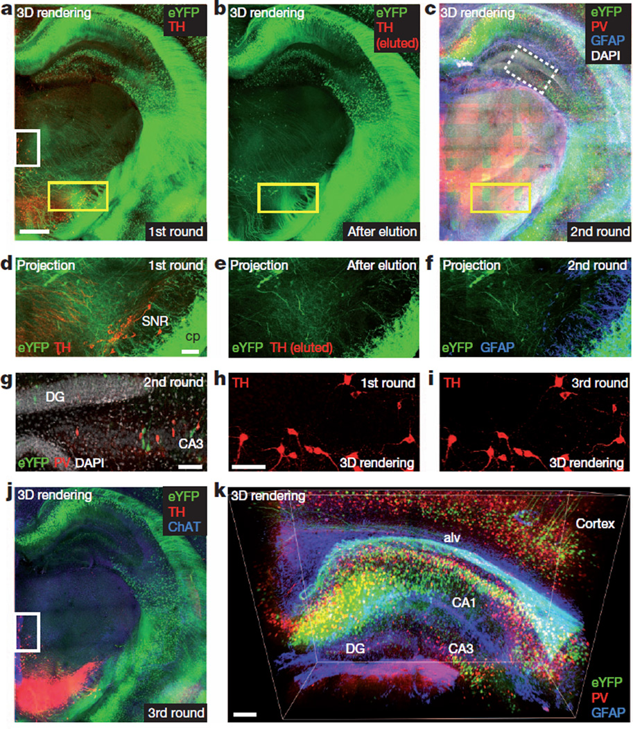
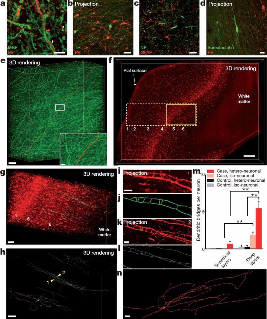
Obtaining high-resolution information from a complex system, while maintaining the global perspective needed to understand system function, represents a key challenge in biology. Here we address this challenge with a method (termed CLARITY) for the transformation of intact tissue into a nanoporous hydrogel-hybridized form (crosslinked to a three-dimensional network of hydrophilic polymers) that is fully assembled but optically transparent and macromolecule-permeable. Using mouse brains, we show intact-tissue imaging of long-range projections, local circuit wiring, cellular relationships, subcellular structures, protein complexes, nucleic acids and neurotransmitters. CLARITY also enables intact-tissue in situ hybridization, immunohistochemistry with multiple rounds of staining and de-staining in non-sectioned tissue, and antibody labelling throughout the intact adult mouse brain. Finally, we show that CLARITY enables fine structural analysis of clinical samples, including non-sectioned human tissue from a neuropsychiatric-disease setting, establishing a path for the transmutation of human tissue into a stable, intact and accessible form suitable for probing structural and molecular underpinnings of physiological function and disease.


Figures

Figure 1
Figure 1. CLARITY
Tissue is crosslinked with formaldehyde (red) in the presence of hydrogel monomers (blue), covalently linking tissue elements to monomers that are then polymerized into a hydrogel mesh (followed by a day-4 wash step; Methods). Electric fields applied across the sample in ionic detergent actively transport micelles into, and lipids out of, the tissue, leaving fine-structure and crosslinked biomolecules in place. The ETC chamber is depicted in the boxed region (Supplementary Fig. 2).
Figure 2
Figure 2. Intact adult mouse brain imaging
Imaging was performed in adult mouse brains (3 months old). a, Cajal quote before CLARITY. b, Cajal quote after CLARITY: Thy1–eYFP line-H mouse brain after hydrogel–tissue hybridization, ETC and refractive-index matching (Methods). c, Fluorescence image of brain depicted in b. d, Dorsal aspect is imaged (single-photon (1p) microscopy), then brain is inverted and ventral aspect imaged. e, Three-dimensional rendering of clarified brain imaged (×10 water-immersion objective; numerical aperture, 0.3; working distance, 3.6 mm). Left, dorsal half (stack size, 3,100 µm; step size, 20 µm). Right, ventral half (stack size, 3,400 µm; step size, 20 µm). Scale bar, 1 mm (Supplementary Videos 3–5). f, Non-sectioned mouse brain tissue showing cortex, hippocampus and thalamus (×10 objective; stack size, 3,400 µm; step size, 2 µm). Scale bar, 400 µm (Supplementary Video 2). gl, Optical sections from f showing negligible resolution loss even at ~3,400-µm deep: z=446 µm (g, h), z=1,683 µm (i, j) and z=3,384 µm (k, l). h, j and l, boxed regions in g, i and k, respectively. Scale bars, 100 µm.m, Cross-section of axons in clarified Thy1–channelrhodopsin2 (ChR2)–eYFP striatum: membrane-localized ChR2–eYFP (1-mm-thick coronal block; ×63 glycerol-immersion objective; numerical aperture, 1.3; working distance, 280 µm). Scale bar, 5 µm. n, Dendrites and spines of neurons in clarified Thy1–eYFP line-H cortex (1-mm-thick coronal block; ×63 glycerol objective). Scale bar, 5 µm.
Figure 3
Figure 3. Molecular phenotyping in intact tissue
a, Protein loss in clarified mouse brain compared to conventional methods (see Supplementary Information for more details); error bars denote s.e.m.; n=4 for each condition. b, Rendering of a 1-mm-thick non-sectioned coronal block of Thy1–eYFP mouse brain immunostained for GFP. The tissue was ETC-cleared (1 day), immunostained (3 days) and imaged (×10 water-immersion objective; single-photon excitation). Left, eYFP (green); middle, anti-GFP (red); right, overlay. Scale bar, 500 µm (Supplementary Video 6). c, Three-dimensional rendering of the boxed region in the cortex in b shows eYFP fluorescence (left) and anti-GFP staining (right). d, Left, co-localization: Manders overlap coefficient plotted versus depth. Right, optical sections at different depths in three-dimensional rendering. Scale bar, 100 µm. e, f, 500-µm-thick block of line-H mouse brain (2 months old) clarified for 1 day and immunostained for synapsin I (red) and PSD-95 (blue) for 3 days (Methods) (×63 glycerol objective; single-photon excitation). e, Left, optical sections (z=20 µm, z=200 µm). Right, enlarged images of boxed regions on left. Individual synaptic puncta resolved throughout depth. White depicts eYFP staining. f, Average immunofluorescence cross-section of PSD-95 puncta at z=20 µm (top) and z=200 µm (bottom). g, Full width at half maximum (FWHM) of average immunofluorescence cross-section of PSD-95 puncta versus depth. Insets, average puncta at z=20 µm and z=200 µm. h, Hippocampal staining. Left, GABA; middle, parvalbumin (PV); right, overlay. 500-µm-thick block of wild-type mouse brain (3 months) clarified (1 day) and immunostained (3 days) (×25 water-immersion objective; numerical aperture, 0.95; working distance, 2.5 mm; single-photon excitation). Scale bar, 20 µm. i, in situ hybridization. Clarified 500-µm mouse brain block showing dopamine receptor D2 (Drd2) mRNA in the striatum. LV, lateral ventricle. Blue, DAPI. 50-base-pair RNA probes for Drd2 visualized with FastRed (×25 water-immersion objective; single-photon excitation (555 nm) for FastRed, two-photon excitation (720 nm) for DAPI). Scale bars: left, 100 µm; right, 20 µm. j, k, Axonal fibres of tyrosine hydroxylase (TH)-positive neurons in the nucleus accumbens (NAc) and caudate–putamen (CPu). j, Three-dimensional rendering of 1-mm-thick clarified mouse brain block stained for tyrosine hydroxylase (red) and DAPI (green). aca, anterior commissure. Scale bar, 500 µm. k, Maximum projection, NAc/aca volume in j. Scale bar, 50 µm.
Figure 4
Figure 4. Multi-round molecular phenotyping of intact tissue
a, First round. Rendering of 1-mm-thick Thy1–eYFP block immunostained for tyrosine hydroxylase in non-sectioned form. ETC-cleared (1 day) and immunostained (6 days). Scale bar, 500 µm (Supplementary Video 10). b, Antibodies eluted from block in a (4% SDS, 60 °C for 0.5 days). Tyrosine hydroxylase signal was removed and eYFP fluorescence retained (Supplementary Video 11). c, Second round. Three-dimensional rendering of same block now immunostained for parvalbumin (red), glial fribrillary acidic protein (GFAP) (blue) and DAPI (white) (Supplementary Video 12). df, Maximum projections of 100 µm volume of yellow-boxed regions in a, b and c, respectively. eYFP-positive neurons preserved. cp, cerebral peduncle; SNR, substantia nigra. Scale bar, 100 µm. g, Optical section of white/dotted-box region in c showing DAPI. CA, cornu ammonis; DG, dentate gyrus. Scale bar, 100 µm. h, i, Tyrosine hydroxylase channel of white box regions in a (h) and j (i). Tyrosine hydroxylase antigenicity preserved through multiple elutions. Scale bar, 100 µm. j, Third round. Block in ac immunostained for tyrosine hydroxylase (red) and choline acetyltransferase (ChAT) (blue) (Supplementary Video 13). k, Three-dimensional view of hippocampus in c showing eYFP-expressing neurons (green), parvalbumin-positive neurons (red) and GFAP (blue). Alv, alveus. Scale bar, 200 µm (Supplementary Video 14).
Figure 5
Figure 5. Human brain structural/molecular phenotyping
Human BA10 500-µm-thick intact blocks clarified (1 day) and immunostained (3 days) (×25 water-immersion objective). a, Optical section: myelin basic protein (MBP) and parvalbumin staining. White arrowheads indicate membrane-localized myelin basic protein around parvalbumin-positive projections. Scale bar, 10 µm. b, Tyrosine hydroxylase and parvalbumin staining (maximum projection; 120 µm volume; step size, 0.5 µm). Scale bar, 50 µm. c, Optical section: neurofilament (NP) and GFAP. Scale bar, 20 µm. d, Somatostatin and parvalbumin staining (maximum projection; 63 µm volume; step size, 0.5 µm). Scale bar, 20 µm. e, Rendering of neurofilament-positive axonal fibres. Red, traced axon across volume. Scale bar, 500 µm. Inset: boxed region. Scale bar, 20 µm (Supplementary Video 15). f, Visualization of parvalbumin-positive neurons in the neocortex of autism case; layers identified as described in ref. 44. Scale bar, 500 µm (Supplementary Video 16). g, Yellow-boxed region in f showing parvalbumin-positive cell bodies and fibres in layers 4, 5 and 6. Three representative parvalbumin-positive interneurons in layer 6 with ladder-shaped hetero- or iso-neuronal connections were traced (green, purple, blue). Scale bar, 100 µm (Supplementary Video 17). h, Three-dimensional rendering of abnormal neurons in g; yellow arrowheads (1, 2) indicate ladder-shaped structures shown below in i and k. Scale bar, 80 µm. i, Zoomed-in maximum projection of 8 µm volume showing morphology of ladder-shaped structure formed by neurites from a single neuron. Scale bar, 10 µm. j, Tracing of structure in i. k, Maximum projection of 18 µmvolume showing ladder-shaped structure formed by neurites from two different neurons. Scale bar, 10 µm. l, Tracing of structure in k. m, Iso- and hetero-neuronal dendritic bridges per neuron. Neurons selected randomly and traced in software (Methods); dendritic bridges were manually counted. **P<0.05; error bars denote s.e.m. n=6 neurons for both superficial and deep layers of autism case and n=4 neurons for both superficial and deep layers of control case. n, Three-dimensional reconstruction of a neuron in layer 2 (superficial) of the autism case. Typical avoidance of iso-dendritic contact was observed.

Comment in

  • CLARITY--a clearer view of the brain.
    Pointer K, Kuo JS, Dempsey RJ. Pointer K, et al. Neurosurgery. 2013 Aug;73(2):N16. doi: 10.1227/01.neu.0000432622.44397.74. Neurosurgery. 2013. PMID: 23867270 No abstract available.
  • CLARITY or the invisible brain.
    Jaber M. Jaber M. Mov Disord. 2013 Oct;28(12):1639. doi: 10.1002/mds.25619. Epub 2013 Sep 16. Mov Disord. 2013. PMID: 24105932 No abstract available.

References

    1. Denk W, Strickler JH, Webb WW. Two-photon laser scanning fluorescence microscopy. Science. 1990;248:73–76. - PubMed
    1. Carmeliet P, Tessier-Lavigne M. Common mechanisms of nerve and blood vessel wiring. Nature. 2005;436:193–200. - PubMed
    1. Helmchen F, Denk W. Deep tissue two-photon microscopy. Nature Methods. 2005;2:932–940. - PubMed
    1. Denk W, Horstmann H. Serial block-face scanning electron microscopy to reconstruct three-dimensional tissue nanostructure. PLoS Biol. 2004;2:e329. - PMC - PubMed
    1. Micheva KD, Smith SJ. Array tomography: a new tool for imaging the molecular architecture and ultrastructure of neural circuits. Neuron. 2007;55:25–36. - PMC - PubMed

Publication types