Anatomical changes in occipitalization: is there an increased risk during the standard posterior approach?
- PMID: 23575658
- PMCID: PMC3641254
- DOI: 10.1007/s00586-013-2768-7
Anatomical changes in occipitalization: is there an increased risk during the standard posterior approach?
Abstract
Purpose: The purpose of this study was to examine the anatomic changes in a case of occipitalization of the atlas.
Methods: Occipitalization of the atlas was found accidentally in a 64-year-old male cadaver. Anatomic dissection was carried out to examine the posterior aspect of the upper cervical region and craniocervical junction. The occipitalized atlas was then harvested and macerated to study the bony anomaly.
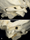
Results: In this case of occipitalization, fusion was observed at both lateral masses and at the left posterior hemiarch of the atlas. We found the following soft tissue changes: the rectus capitis posterior minor muscle was lacking on the left side and was atrophic on the right, the obliquus capitis superior muscle was present on both sides showing moderate atrophy and fatty changes. The posterior atlanto-axial membrane was thinner and asymmetric, had a free edge on the right side. Lateral to this edge the dura was lying free. We believe that these changes of the posterior atlanto-axial membrane together with the increased distance between the fused posterior arch of the atlas and the lamina of the axis could cause the observed "dura bulge" at this level. The vertebral artery was entering the skull through a canal on the left side.
Conclusions: In our case, occipitalization considerably changed the anatomy of the upper cervical spine and craniocervical junction. Special care must be taken when using the posterior approach to avoid neurovascular injury in cases with occipitalization.
Figures


References
-
- Hensinger RN (1989) Anomalies of the atlas. In: The cervical spine research society, the cervical spine, 2nd edn. J.B. Lippincott Company, Philadelphia, pp 244–247
-
- Dieckmann H. Basilare impression. Hippokrates Verlag Stuttgart: Atlasassimilation und andere Skelettfehlbildungen der Zerviko-okzipital-Region; 1966.
-
- von Torklus D, Gehle W (1970) Die obere Halswirbelsäule. Georg Thieme Verlag Stuttgart
Publication types
MeSH terms
LinkOut - more resources
Full Text Sources
Other Literature Sources

