. 2013:4:1662.
doi: 10.1038/ncomms2677.

FBXW7α attenuates inflammatory signalling by downregulating C/EBPδ and its target gene Tlr4

Affiliations

FBXW7α attenuates inflammatory signalling by downregulating C/EBPδ and its target gene Tlr4

Kuppusamy Balamurugan et al. Nat Commun. 2013.

Abstract

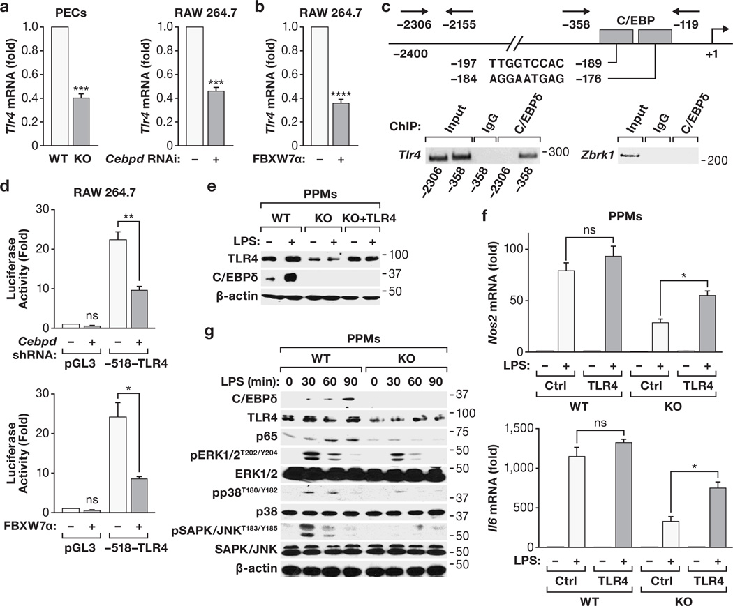
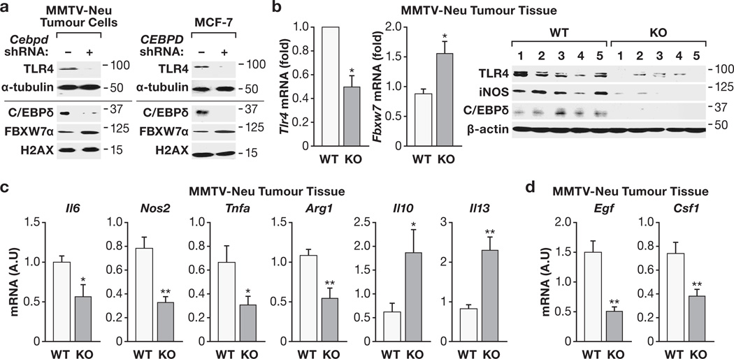
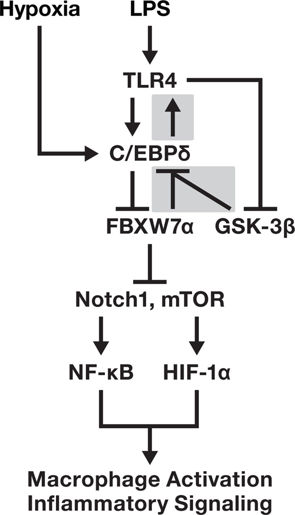
Toll-like receptor 4 (Tlr4) has a pivotal role in innate immune responses, and the transcription factor CCAAT/enhancer binding protein delta (C/EBPδ, Cebpd) is a Tlr4-induced gene. Here we identify a positive feedback loop in which C/EBPδ activates Tlr4 gene expression in macrophages and tumour cells. In addition, we discovered a negative feedback loop whereby the tumour suppressor FBXW7α (FBW7, Cdc4), whose gene expression is inhibited by C/EBPδ, targets C/EBPδ for degradation when C/EBPδ is phosphorylated by GSK-3β. Consequently, FBXW7α suppresses Tlr4 expression and responses to the ligand lipopolysaccharide. FBXW7α depletion alone is sufficient to augment pro-inflammatory signalling in vivo. Moreover, as inflammatory pathways are known to modulate tumour biology, Cebpd null mammary tumours, which have reduced metastatic potential, show altered expression of inflammation-associated genes. Together, these findings reveal a role for C/EBPδ upstream of Tlr4 signalling and uncover a function for FBXW7α as an attenuator of inflammatory signalling.


Figures

Figure 1
Figure 1. C/EBPδ promotes HIF-1α expression in macrophages through inhibition of FBXW7α
a) RT-PCR analysis of FBXW7 isoform expression from different sources as follows. 1, primary peritoneal macrophages (PPMs); 2, RAW 264.7 cells; 3, MMTV-Neu mammary tumour tissue; 4, primary mouse embryo fibroblasts. Numbers indicate the position of size markers in base pairs. (b) RT-qPCR analysis of Fbxw7 transcript levels in PPMs from WT and Cebpd−/− KO mice, cultured +/− LPS (100 ng/ml, 24 h), compared to WT untreated (n=4, *P<0.05; **P<0.001). (c) Western analysis of nuclear extract (NE) from primary human monocytes nucleofected with siRNA oligos (C, control; D, CEBPD; F, FBXW7) and treated with LPS (100 ng/ml) and 1%O2 (16 h) as indicated. SE, short exposure; LE, long exposure. (d) RT-qPCR analysis of FBXW7 and CEBPD transcripts in primary human monocytes as in panel (c) (n=3, *P<0.05; **P<0.001). (e) Western analysis of NE from PPMs nucelofected with siRNA oligos and treated with LPS (100 ng/ml) and 1%O2 for 16 h as indicated. SE, short exposure; LE, long exposure. Where applicable, data are mean±S.E.M., evaluated by two-tailed unequal variance t-test.
Figure 2
Figure 2. FBXW7α interacts with C/EBPδ and targets it for degradation
(a) RAW 264.7 macrophages were transfected with control or Fbxw7α siRNA oligos for 48 h, then pulse-labeled with 35S-methionine/cysteine, followed by chased with excess unlabeled aminoacids for the indicated times. Quantitation of the phosphorimage of C/EBPδ signal is depicted in the graph (mean±S.E.M., n=2–3) and representative primary data are shown. (b) Western analysis of RAW 264.7 cells transfected with HA-ubiquitin expression constructs and control or Fbxw7α siRNA oligos and treated ± MG132 (20 µM, 6 h), followed by immunoprecipitation under denatured conditions with anti-C/EBPδ or IgG (with equal aliquots of the indicated extracts). Input (2.5% of lysate) was analyzed as indicated. (c) Alignment of phosphodegron motifs present in known FBXW7 substrates with C/EBPδ and its TTS/AAA mutant. (d) Western analysis of RAW 264.7 cells transfected with WT- or TTS/AAA-CEBPδ expression plasmids and/or HA-FBXW7α. (e) Western analysis of RAW 264.7 cells transfected with WT or TTS/AAA C/EBPδ expression plasmids and immunoprecipitated with anti-C/EBPδ or IgG and input (2.5% of lysate) as indicated. (f) RAW 264.7 cells were transfected with WT- or TTS/AAA-C/EBPδ expression constructs. C/EBPδ was immunoprecipitated and the beads were incubated with FBXW7α and HA-ubiquitin as indicated (see Methods for details) and analyzed by Western with anti-HA and C/EBPδ antibodies. (g) Western analysis of RAW 264.7 cells transfected with WT- or TTS/AAA-C/EBPδ, treated ± MG132 (20 µM, 6 h) followed by immunoprecipitation under denatured conditions using anti-C/EBPδ or IgG and input (2.5% of lysate) as indicated. (h) Western analysis of RAW 264.7 cells transfected with WT- and TTS/DDD-C/EBP™ for 48 h followed by immunoprecipitation with anti-C/EBPδ or IgG and input (2.5% of lysate) as indicated. (i) Western analysis (bottom panel) of RAW 264.7 cells transfected with WT-, TTS/AAA- or TTS/DDD-C/EBPδ expression constructs and treated with CHX as indicated. Quantification (top panel) of the C/EBPδ signal was normalized to actin (n=3, *P<0.05; ***P<0.0001; ns, not significant). (j) Western analysis of RAW 264.7 cells transfected with WT- or TTS/DDD-C/EBPδ expression constructs and treated ± MG132 (20 µM, 6 h). Where applicable, data are mean±S.E.M., evaluated by two-tailed unequal variance t-test.
Figure 3
Figure 3. C/EBPδ stability is regulated by GSK-3β phosphorylation
(a) RAW 264.7 cells were transfected with WT or TTS/AAA CEBPδ expression plasmids and treated ± GSK-3β inhibitor CHIR99021 (5 µM, 2 h). Cell extracts were immunoprecipitated with anti-C/EBPδ or IgG (with an aliquot of the indicated extract) and the Western analyzed with anti-phosphothreonine (pT) antibody. (b) Western analysis of NE from PPMs (left panel) or RAW 264.7 cells (right panel) treated with GSK-3β inhibitors CHIR99021 or BIO (6-bromoindirubin-3’-oxime) (5 µM, 2 h). β-catenin, which is known to be targeted for degradation by GSK-3β phosphorylation, served as positive control. (c) Western analysis of RAW 264.7 cells transfected with WT- or TTS/DDD-C/EBPδ expression plasmids and treated ± GSK-3β inhibitor CHIR99021 for 2 h. (d) GSK-3β phosphorylates C/EBPδ in vitro. HEK293T cells were transfected with WT- or TTS/AAA-C/EBPδ expression plasmids. C/EBPδ proteins were immunoprecipitated from cell extracts and in vitro kinase assay reactions were carried out in the presence or absence of GSK-3β. Samples were resolved by SDS-PAGE and subjected to autoradiography (top panel). Total radioactivity (bottom panel) incorporated into the C/EBPδ protein was quantified (n=3). Representative input levels of C/EBPδ are shown by Western (inset). (e) RAW 264.7 cells were treated ± LPS (4 h) and cell extracts were immunoprecipitated with anti-C/EBPδ or IgG antibodies and Western analysis was carried out with anti-phosphothreonine (pT) antibody. Input (2.5% of the lysate) was analyzed as indicated. (f) Western analysis (top panel) of RAW 264.7 cells treated ± LPS (100 ng/ml, 4 h) followed by CHX for the indicated times, and quantification of C/EBPδ normalized to β-actin signal (bottom graph) compared to respective untreated (n=3, *P<0.05; **P<0.001; ****P <0.0001). (g) Western analysis of RAW 264.7 cells transfected with HA-GSK-3β-S9A expression plasmids treated ± LPS (4 h) as indicated. (h) Western analysis of RAW 264.7 cells pretreated with the PI3K/AKT kinase pathway inhibitor LY294002 (10 µM, 1 h) followed by LPS (100 ng/ml, 4 h) as indicated. Where applicable, data are mean±S.E.M., evaluated by two-tailed unequal variance t-test.
Figure 4
Figure 4. FBXW7α suppresses TLR4-mediated LPS responses through C/EBPδ
(a) Western analysis of NE from PPMs transfected with vector or FBXW7α expression constructs and treated with LPS (100 ng/ml, 16 h) as indicated. (b) RT-qPCR of the indicated mRNA levels in PPMs nucelofected with FBXW7α expression plasmids and treated with LPS (100 ng/ml, 16 h) as indicated (n=3, * P<0.05, **P<0.001, ****P<0.0001). (c) Western analysis of PPMs transfected with HA-FBXW7α and treated with LPS (100 ng/ml, 16h) as indicated. SE, short exposure; LE, long exposure. (d) Western analysis of proteins from PPMs nucleofected with control or Fbxw7α siRNA oligos and treated ± LPS (100 ng/ml, 16 h). The line separates analysis of cytoplasmic (top) and nuclear (bottom) extracts. (e) Western analysis of RAW 264.7 macrophages transfected with siRNA oligos against endogenous Cebpd or Fbxw7α alone or in combination and treated with LPS (100 ng/ml, 16 h) as indicated. The line separates analysis of cytoplasmic (top) and nuclear (bottom) extracts. (f) Western analysis of RAW 264.7 macrophages transfected with the indicated expression plasmids. (g) Western analysis of PECs isolated from FVB/N mice 48 h after injection of RNAi against Fbxw7α. or control siRNA. (h) Plasma IL-6 concentrations from mice injected with RNAi against Fbxw7α- or control siRNA for 72 h followed by LPS (40 ng) or vehicle (saline) for 1 h. The horizontal line indicates the median IL-6 concentration (n=4–5, P values were determined by the Wilcoxon rank-sum test, P < 0.01). (i) Western analysis of PPMs transfected with control or Fbxw7α siRNA oligos and treated with LPS (100 ng/ml, 4 h) as indicated. SE, LE; short, long exposure. Where applicable, data are mean±S.E.M., evaluated by two-tailed unequal variance t-test (except panel 4h).
Figure 5
Figure 5. TLR4 is a direct transcriptional target of C/EBPδ
(a) RT-qPCR analysis of Tlr4 mRNA levels in WT or Cebpd KO PECs and in RAW 264.7 macrophages after Cebpd silencing (n=3, ***P<0.001). (b) RT-qPCR analysis of Tlr4 in RAW 264.7 macrophages with or without ectopic FBXW7α (n=3, ****P<0.0001). (c) Schematic of the Tlr4 promoter with location of putative C/EBP binding sites and of primers used for ChIP. ChIP analysis from PPMs for binding of C/EBPδ to the indicated Tlr4 promoter regions. IgG and a Zbrk1 promoter region served as negative controls. Numbers indicate the position of molecular size markers in base pairs. (d) Luciferase reporter assay in RAW 264.7 macrophages transfected with the indicated luciferase reporter and shRNA against Cebpd (top panel) or FBXW7α expression constructs (bottom panel) (n=3, *P<0.05, **P<0.001). ns, not significant. (e) Western analysis of PPMs from WT and Cebpd KO mice transfected with TLR4 expression plasmids as indicated. (f) RT-qPCR analysis of the indicated mRNA levels in PPMs as in panel (e). (n=3, *P<0.05; ns, not significant). (g) Western analysis of PPMs from WT and Cebpd KO mice treated with 200 ng/ml LPS for the indicated times. Where applicable, data are mean±S.E.M., evaluated by two-tailed unequal variance t-test.
Figure 6
Figure 6. Cebpd null tumours exhibit reduced expression of TLR4 and altered expression of inflammatory genes
(a) Western analysis of proteins from an MMTV-Neu tumour cell line or MCF-7 cells with stable shRNA-silencing of C/EBP™ expression or control shRNA. The line separates analysis of cytoplasmic (top) and nuclear (bottom) extracts. (b) RT-qPCR analysis of Tlr4 and Fbxw7 mRNA (left panels) and Western analysis (right panel) of protein from MMTV-Neu tumours of WT and Cebpd KO mice (n=5, *P<0.05). (c,d) RT-qPCR analysis of mRNA levels of the indicated genes in MMTV-Neu tumour tissue from WT and Cebpd KO mice. A.U., arbitrary units. (n=6, *P<0.05; ** P<0.001). Where applicable, data are mean±S.E.M., evaluated by two-tailed unequal variance t-test.
Figure 7
Figure 7. Schematic describing the feedback loops between TLR4, C/EBPδ and FBXW7α that control LPS signaling
The shaded boxes indicate the elements of this pathway that were identified in this study.

Similar articles

Cited by

References

    1. Kawai T, Akira S. Toll-like receptors and their crosstalk with other innate receptors in infection and immunity. Immunity. 2011;34:637–650. - PubMed
    1. Roger T, et al. Protection from lethal gram-negative bacterial sepsis by targeting Toll-like receptor 4. Proc Natl Acad Sci U S A. 2009;106:2348–2352. - PMC - PubMed
    1. Lin Q, Li M, Fang D, Fang J, Su SB. The essential roles of Toll-like receptor signaling pathways in sterile inflammatory diseases. Int Immunopharmacol. 2011;11:1422–1432. - PubMed
    1. Huang B, et al. Toll-like receptors on tumor cells facilitate evasion of immune surveillance. Cancer Res. 2005;65:5009–5014. - PubMed
    1. Szczepanski MJ, et al. Triggering of Toll-like receptor 4 expressed on human head and neck squamous cell carcinoma promotes tumor development and protects the tumor from immune attack. Cancer Res. 2009;69:3105–3113. - PMC - PubMed

Publication types

MeSH terms

LinkOut - more resources