. 2013:4:1665.
doi: 10.1038/ncomms2647.

Mitotic spindle orientation predicts outer radial glial cell generation in human neocortex

Affiliations

Mitotic spindle orientation predicts outer radial glial cell generation in human neocortex

Bridget E LaMonica et al. Nat Commun. 2013.

Abstract

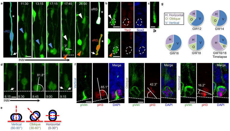
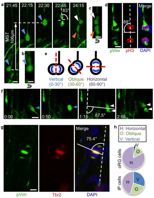
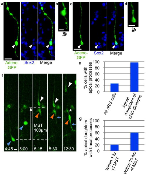
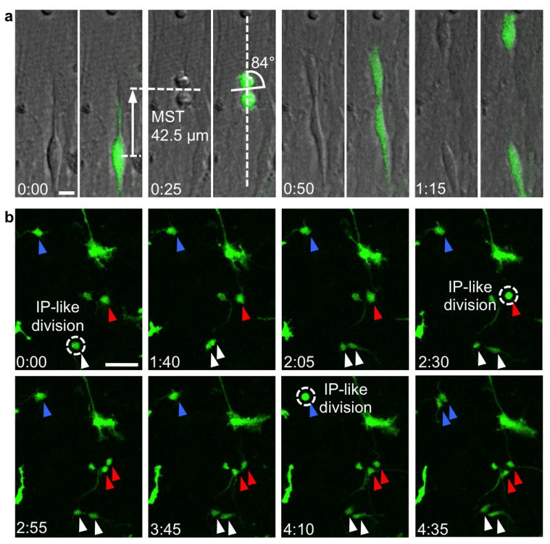
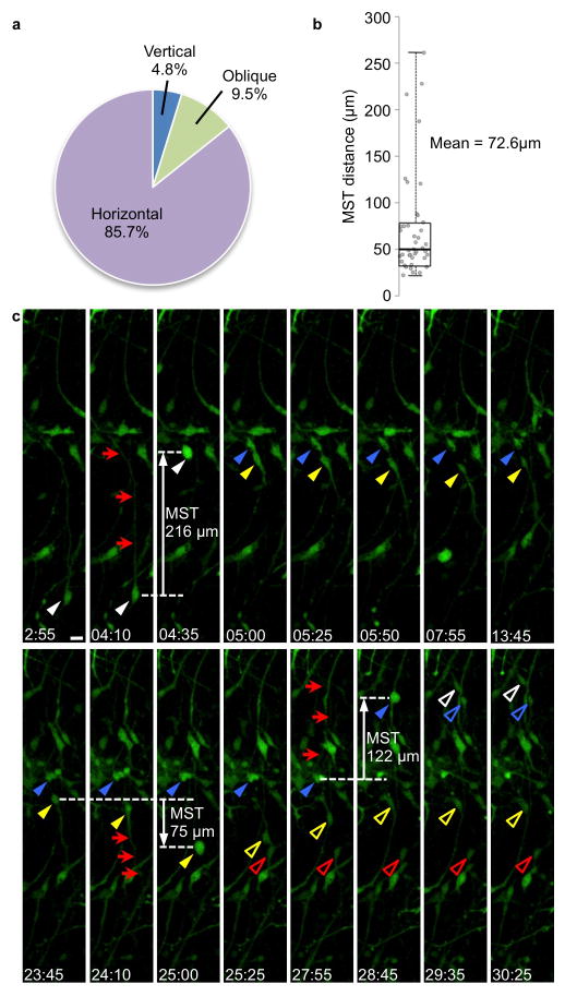
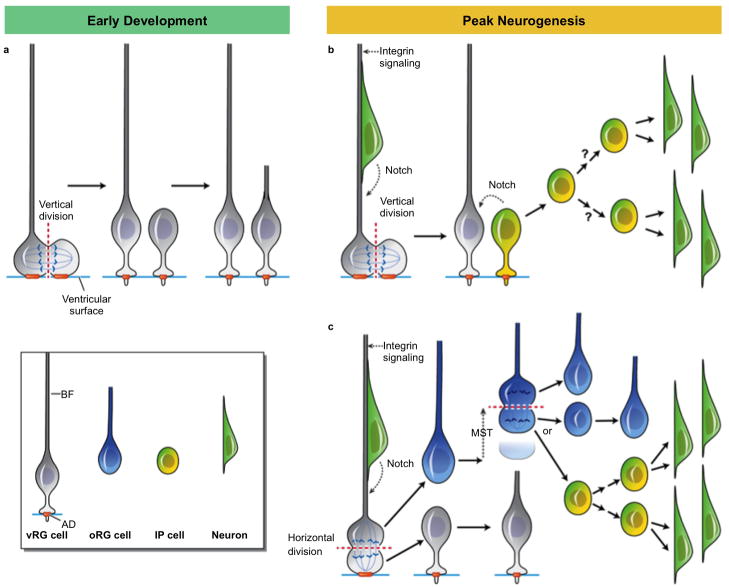
The human neocortex is increased in size and complexity as compared with most other species. Neocortical expansion has recently been attributed to protracted neurogenesis by outer radial glial cells in the outer subventricular zone, a region present in humans but not in rodents. The mechanisms of human outer radial glial cell generation are unknown, but are proposed to involve division of ventricular radial glial cells; neural stem cells present in all developing mammals. Here we show that human ventricular radial glial cells produce outer radial glial cells and seed formation of the outer subventricular zone via horizontal divisions, which occur more frequently in humans than in rodents. We further find that outer radial glial cell mitotic behaviour is cell intrinsic, and that the basal fibre, inherited by outer radial glial cells after ventricular radial glial division, determines cleavage angle. Our results suggest that altered regulation of mitotic spindle orientation increased outer radial glial cell number, and ultimately neuronal number, during human brain evolution.


Figures

Figure 1
Figure 1. Horizontal vRG divisions produce oRG cells
a) Time-lapse stills of horizontal vRG division labeled with Adeno-GFP in GW16 human fetal cortical slice that results in generation of an oRG cell. vRG cell (blue arrowhead) starts at a distance from the ventricle, descends during interkinetic nuclear migration, and divides with a horizontal spindle orientation at the ventricular surface (dotted white line at 17:45). Basal daughter (white arrowhead) retains basal fiber (asterisk) and rapidly exits the ventricular zone, adopting oRG cell morphology. Apical daughter (orange arrowhead) inherits entire apical domain (white arrow) and regrows a basal fiber, becoming a self-renewed vRG cell. Cells of interest are schematized at beginning and end of sequence to more clearly demonstrate morphology and inheritance of basal fiber and apical domain. Scale bar, 10μm. b) Fate staining of daughter cells from division in (a) confirms that the basal daughter (white arrowhead) is Tbr2−/Sox2+, consistent with oRG identity, and the apical daughter (orange arrowhead) is also Tbr2−/Sox2+, consistent with vRG identity. Scale bar, 10μm. c) Z-plane analysis of basal daughter from division in (a) rotated 90° confirms lack of apical process. Scale bar, 10μm. d) Time-lapse stills of vertical vRG division in GW16 human fetal cortical slice after which both daughters inherit half of the AD (white arrows). and neither daughter adopts oRG morphology. Part of the basal fiber is not visible due to its location out of the imaged z-stack. Scale bar, 10μm. e) Schematic of vRG cells undergoing mitosis at the ventricular surface with vertical, oblique, and horizontal cleavage angles. Mitotic spindle orientation was analyzed with respect to the ventricular surface. f) Images of vRG cells in fixed human fetal cortical slices undergoing mitosis with vertical (left), oblique (center), and horizontal (right) mitotic spindle orientations at the ventricular surface. Scale bars, 5μm. g) Quantification of mitotic spindle orientation in fixed slices throughout peak neurogenesis and oRG cell generation, and during time-lapse imaging of GW16/18 human fetal cortical slices. Cells used for calculation in fixed slices were pH3+, pVim+ cells in anaphase or telophase that displayed a basal fiber and were located at the ventricular surface.
Figure 2
Figure 2. oRG cells divide horizontally to self-renew
a) Time-lapse stills of a dividing oRG cell (blue arrowhead) in GW16 human fetal cortical slice labeled with Adeno-GFP. The cell undergoes MST and divides with a horizontal spindle orientation, producing an apical daughter (orange arrowhead) and a basal daughter (white arrowhead). The basal daughter retains the basal fiber and becomes a self-renewed oRG cell, while the apical daughter produces an apical process. Scale bar 10μm. b) Z-plane analysis of oRG cell in first frame of (a), demonstrating lack of apical process. Scale bar, 10μm. c) Z-plane analysis of daughter cells in last frame of (a), demonstrating lack of apical process on basal daughter, and presence of apical process on apical daughter. Scale bar, 10μm. d) Image of oRG cell in fixed GW16 human fetal cortical oSVZ undergoing mitosis with a horizontal cleavage plane. Scale bar, 10μm. e) Schematic of oRG cells undergoing mitosis with vertical, oblique, and horizontal cleavage planes. Cleavage plane angle was analyzed with respect to the basal fiber of the dividing cell (or in the case of IP cells, with respect to the nearest basal fiber). f) Time-lapse stills of IP cell (blue arrowhead) in a GW18 human fetal cortical slice that retracts its processes and rounds up to divide symmetrically, without undergoing MST or interkinetic nuclear migration, to produce two daughter cells (white arrowheads). Spindle orientation was calculated with respect to the nearest radial fiber. Scale bar, 20μm. g) Image of IP cell in fixed GW16 human fetal cortical oSVZ undergoing mitosis with a horizontal spindle orientation. Scale bar, 5μm. h) Quantification of mitotic spindle orientations in i/oSVZ progenitors (oRG cells and IP cells) in fixed GW16 human fetal cortical slices. Cells used for calculation were in anaphase or telophase and displayed the following characteristics: oRG cells: pVim+, pH3+ cells in the iSVZ or oSVZ that displayed a basal fiber but no apical ventricular contact; IP cells: Tbr2+ cells in the iSVZ or oSVZ that were pVim+ and displayed no basal or apical fiber.
Figure 3
Figure 3. Apical and basal processes on oRG cells and oRG apical daughters
a) oRG cell with apical process in GW18 human fetal oSVZ, identified by morphology after 18-hour Adeno-GFP infection followed by fixation and Sox2 staining. Scale bar, 20 μm. b) Z-plane analysis of cell in (a) rotated 90° confirms presence of apical process. Scale bar, 10μm. c) oRG cell lacking apical process in GW18 human fetal oSVZ, identified by morphology after Adeno-GFP infection and by Sox2 positivity. Scale bar, 20μm. d) Z-plane analysis of cell in (c) rotated 90° confirms lack of apical process. Scale bar, 10μm. e) Quantification of presence of apical process on all oRG cells in the GW16/18 human fetal i/oSVZ, and on apical daughters of oRG cell divisions after time-lapse imaging of GW16/18 human fetal cortex. All apical processes on apical daughters were produced within 1 hour of MST. f) Time-lapse stills of a dividing oRG cell (blue arrowhead) in GW18 human fetal cortical slice labeled with Adeno-GFP. The oRG cell undergoes MST and produces an apical daughter (orange arrowhead) and a basal daughter (white arrowhead). The basal daughter retains the basal fiber and becomes a self-renewed oRG cell. The apical daughter produces an apical process immediately after MST (blue arrow), and a basal process several hours after MST (asterisk), adopting bipolar morphology. Scale bar 10μm. g) Quantification of presence of basal process on apical daughters of oRG cell divisions after time-lapse imaging in GW16/18 human fetal cortex. Unlike the apical process, the basal process often takes more than 1 hour to begin growing after MST.
Figure 4
Figure 4. Dissociated cortical progenitor cells display MST and horizontal spindle orientation
a) oRG-like cell in dissociated culture undergoes MST followed by division with horizontal cleavage angle. Dissociated cortical cells were infected with low-titer GFP adenovirus or retrovirus and grown in culture for up to 5 weeks. Transmitted light microscopy allowed visualization of condensed chromatin for quantification of spindle orientation. Scale bar 10μm. b) Multiple IP-like cell divisions in culture, which contrast with oRG-like divisions by their lack of MST. Individual multipolar cells were followed (blue, red, and white arrowheads), and over a period of less than 5 hours, each cell underwent mitosis (dotted circle) without MST, producing two multipolar daughter cells. Scale bar 50 μm.
Figure 5
Figure 5. MST and establishment of a horizontal spindle orientation are cell-intrinsic processes
a)Quantification of cleavage angles in oRG-like cells in dissociated culture in which transmitted light microscopy allowed visualization of spindle orientation. b) Box plot of MST distances for oRG-like divisions in dissociated culture. c) Time-lapse stills of dissociated, Adeno-GFP labeled GW16 human neocortical progenitor cell divisions. oRG-like cell (white arrowhead) with basal process (red arrows) undergoes MST along the basal process and divides at 4:35 to produce two daughter cells: a “basal” daughter (blue arrowhead) and an “apical” daughter (yellow arrowhead). The apical daughter grows a basal fiber (red arrows) in the opposite direction as the original basal fiber, and subsequently undergoes MST in the direction of the newly-generated fiber at 25:00 to produce two daughter cells (open yellow arrowhead, open red arrowhead). The basal daughter of the first division extends its basal fiber (red arrows) in the same orientation as the original basal fiber, and undergoes MST in the direction of this fiber. This cell divides at 28:45 to produce two daughter cells (open white arrowhead, open blue arrowhead). Scale bar, 20 μm.
Figure 6
Figure 6. Model of cleavage angle regulation of cell fate during human cortical developent
a) During early development, vRG divisions at the VZ are likely vertical, resulting in one daughter inheriting the basal fiber (BF), and both daughters inheriting half of the apical domain (AD), orange. The daughter that retains the basal fiber maintains its apical contact and becomes a self-renewed vRG cell, while the daughter that does not receive the basal fiber likely also maintains its apical contact, generates a new basal fiber, and adopts a vRG cell identity, thereby increasing the vRG progenitor population. b) During peak neurogenesis, vRG divisions can either be vertical or horizontal/oblique. vRG division with vertical cleavage plane results in both daughter cells inheriting half of the AD. The cell that retains the basal fiber receives Notch and Integrin signaling via the basal fiber, maintains its apical contact and becomes a self-renewed vRG cell. The cell that does not inherit the basal fiber begins to express IP cell markers, including Notch ligands, thereby contributing to Notch activation of neighboring vRG cells. The newly-generated IP cell delaminates from the apical adhesion belt during the next cell cycle and migrates in a basal direction, where it may undergo multiple rounds of neurogenic divisions. c) vRG division with a horizontal cleavage plane results in the more apical daughter inheriting the entire AD, regrowing a basal fiber, and becoming a self-renewed vRG cell. The more basal daughter inherits the basal fiber, rapidly exits the VZ, and adopts an oRG cell fate. The newly-generated oRG cell continues to divide horizontally, with the basal daughter inheriting the basal fiber and maintaining oRG cell identity. The apical daughter of horizontal oRG cell divisions either regrows a new fiber and adopts oRG cell fate, expanding the oRG cell population, or becomes a IP cell and undergoes multiple rounds of neurogenic divisions.

References

    1. Lui JH, Hansen DV, Kriegstein AR. Development and evolution of the human neocortex. Cell. 2011;146:18–36. - PMC - PubMed
    1. Lamonica BE, Lui JH, Wang X, Kriegstein AR. OSVZ progenitors in the human cortex: an updated perspective on neurodevelopmental disease. Curr Opin Neurobiol. 2012 - PMC - PubMed
    1. Hansen DV, Lui JH, Parker PR, Kriegstein AR. Neurogenic radial glia in the outer subventricular zone of human neocortex. Nature. 2010;464:554–561. - PubMed
    1. Fietz SA, et al. OSVZ progenitors of human and ferret neocortex are epithelial-like and expand by integrin signaling. Nat Neurosci. 2010;13:690–699. - PubMed
    1. Martinez-Cerdeno V, et al. Comparative analysisof the subventricular zone in rat, ferret and macaque: evidence for an outer subventricular zone in rodents. PLoS One. 2012;7:e30178. - PMC - PubMed

Publication types