. 2013 Apr 10;33(15):6524-39.
doi: 10.1523/JNEUROSCI.3838-12.2013.

Tumor necrosis factor-alpha is produced by dying retinal neurons and is required for Muller glia proliferation during zebrafish retinal regeneration

Affiliations

Tumor necrosis factor-alpha is produced by dying retinal neurons and is required for Muller glia proliferation during zebrafish retinal regeneration

Craig M Nelson et al. J Neurosci. .

Abstract

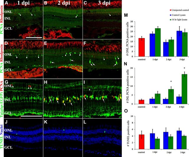
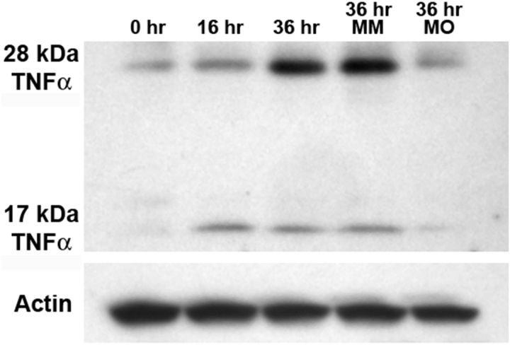
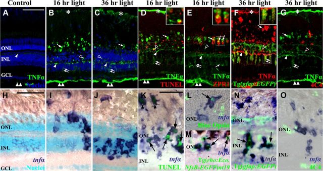
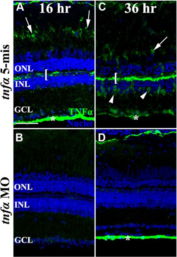
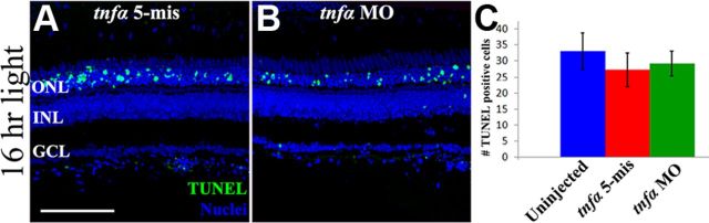
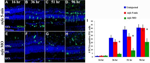
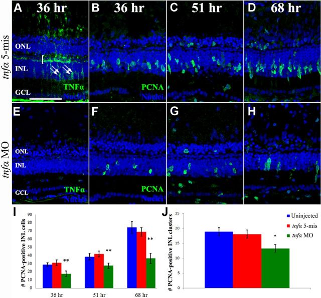
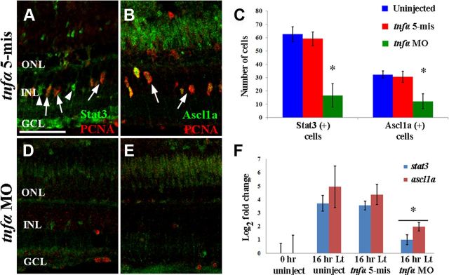
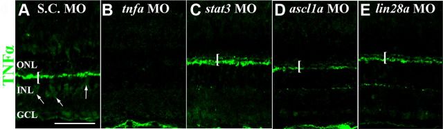
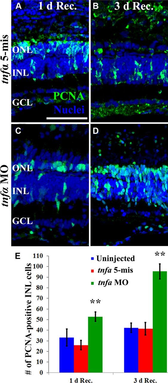
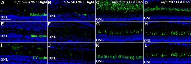
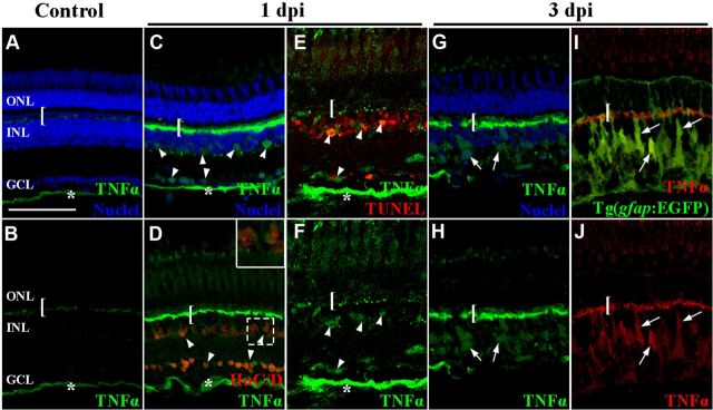
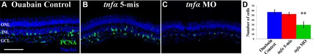
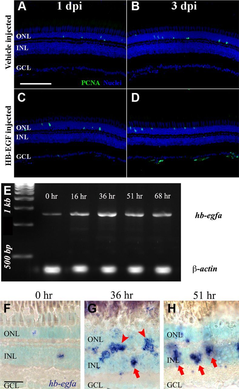
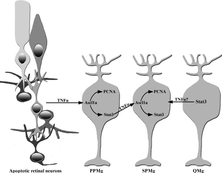
Intense light exposure causes photoreceptor apoptosis in dark-adapted adult albino zebrafish (Danio rerio). Subsequently, Müller glia increase expression of the Achaete-scute complex-like 1a (Ascl1a) and Signal transducer and activator of transcription 3 (Stat3) transcription factors and re-enter the cell cycle to yield undifferentiated neuronal progenitors that continue to proliferate, migrate to the outer nuclear layer, and differentiate into photoreceptors. A proteomic analysis of light-damaged retinal homogenates, which induced Müller glia proliferation when injected into an undamaged eye, revealed increased expression of tumor necrosis factor α (TNFα) signaling proteins relative to undamaged retinal homogenates. TNFα expression initially increased in apoptotic photoreceptors and later in Müller glia. Morpholino-mediated knockdown of TNFα expression before light damage diminished the expression of both Ascl1a and Stat3 in Müller glia and significantly reduced the number of proliferating Müller glia without affecting photoreceptor cell death. Knockdown of TNFα expression in the Müller glia resulted in fewer proliferating Müller glia, suggesting that Müller glial-derived TNFα recruited additional Müller glia to re-enter the cell cycle. While TNFα is required for increased Ascl1a and Stat3 expression, Ascl1a and Stat3 are both necessary for TNFα expression in Müller glia. Apoptotic inner retinal neurons, resulting from intravitreal injection of ouabain, also exhibited increased TNFα expression that was required for Müller glia proliferation. Thus, TNFα is the first molecule identified that is produced by dying retinal neurons and is necessary to induce Müller glia to proliferate in the zebrafish retinal regeneration response.


Figures

Figure 1.
Figure 1.
Light-damaged retinal homogenate contains a factor that induces Müller glia proliferation. Retinal sections from wild-type eyes that were intravitreally injected with a homogenate from either undamaged retinas (A–C) or light-damaged retinas (D–L) were immunolabeled with anti-rhodopsin (red) to visualize rod outer segments and anti-PCNA (green) to detect proliferating cells in the ONL and INL (arrowheads and arrows, respectively) at 1, 2, and 3 dpi (days postinjection, A–F). Tg(gfap:EGFP)nt11 eyes were injected with light-damaged retinal homogenate and immunolabeled with anti-EGFP and anti-PCNA (G–I, green and red, respectively) at 1, 2, and 3 dpi. The number of PCNA-positive cells significantly increased (p < 0.05, n = 10) in the INL of light-damaged homogenate-injected eyes at 2 and 3 dpi relative to the controls (N), but not in the ONL (M). Injection of the light-damaged retinal homogenate did not cause any cell death as measured by the number of TUNEL-positive cells (JL, O). Scale bars: (in A) AF, JL, 50 μm; (in G) GI, 50 μm.
Figure 2.
Figure 2.
TNFα expression increases during constant light treatment. Total protein was isolated from dark-adapted albino retinas after 0 (Control), 16, and 36 h of constant light treatment. Immunoblotting for TNFα expression reveals a 28 kDa band, likely corresponding to the uncleaved form of TNFα. After 16 h of constant light, a 17 kDa band is detected that corresponds to the mature, secreted form of TNFα. Total protein was also isolated from dark-adapted albino dorsal retinas that were injected and electroporated with either the tnfα 5-mis control morpholino (MM) or the tnfα morpholino (MO) and placed in constant intense light for 36 h. While the tnfα 5-mis control retina had approximately the same amount of TNFα as the uninjected light-damaged retina (36 h), the tnfα morphant possessed dramatically less TNFα protein. β-actin expression was detected as a loading control.
Figure 3.
Figure 3.
TNFα is expressed in different cell types during the constant light treatment. Dark-adapted albino zebrafish were exposed to constant intense light for 0 h, 16 h, or 36 h. Cryosections were immunolabeled for TNFα (AE, G, green; F, red) or in situ hybridized with tnfα antisense riboprobe (HO). TNFα antisera weakly labeled the NFL and OPL in undamaged control retinas (A, double arrowhead and arrowhead, respectively). At 16 and 36 h of constant light (B and C, respectively), TNFα expression increased in the OPL (arrowhead), ONL (open arrowhead), photoreceptor outer segments (arrow), INL (double arrow), NFL (double arrowhead), and retinal pigmented epithelium (RPE; asterisk). Following 36 h of constant intense light (C), TNFα antisera labeling in the INL reveals cells with processes that span from the GCL to the ONL (double arrow). At 16 h of light, TNFα colocalized with some TUNEL-positive ONL cells (D, open arrowhead and inset) and ZPR1-positive double cones (E, arrows and inset). At 36 h, TNFα colocalized with EGFP-positive Müller glia (F, double arrow and inset) in the Tg(gfap:EGFP)nt11 transgenic line, but not with 4C4-positive microglia (G, red). In situ hybridization with a tnfα antisense probe (HO, blue) demonstrates increased tnfα expression primarily in the ONL following 16 h of light (I) and then primarily in the INL at 36 h of treatment (J) relative to the undamaged control (H). Colocalization of the tnfα antisense riboprobe with TUNEL-positive rods and cones (K, arrows and arrowheads, respectively), blue opsin-expressing cones (L, arrow), and EGFP-expressing rod photoreceptors in the Tg[rho:Eco.NfsB-EGFP]nt19 transgenic line (M, arrows) occurred at 16 h of light damage. After 36 h of light damage, the EGFP-expressing Müller glia (N, green) in the Tg(gfap:EGFP)nt11 transgenic line expressed tnfα (N, arrows), while the 4C4-expressing microglia (O, green) did not express TNFα. The boxed insets (DF) are magnified 2.9-fold to highlight the colocalization with TNFα. Scale bars: (in A) AG, 50 μm; (in H) HJ, 50 μm; (in K) KO, 50 μm.
Figure 4.
Figure 4.
In vivo knockdown of TNFα during constant light treatment. Dark-adapted albino zebrafish were intravitreally injected and electroporated with either lissamine-tagged tnfα 5-mismatch morpholinos (tnfα 5-mis, A, C) or translation-inhibiting tnfα morpholinos (tnfα MO, B, D) and then exposed to constant intense light for either 16 h (A, B) or 36 h (C, D). The tnfα morphant retinas showed decreased intensity of TNFα immunolabeling relative to both the uninjected (data not shown) and tnfα 5-mis controls, in the outer segments (arrows), OPL (bracket), Müller glia (arrowheads), and the NFL (asterisk). Scale bar: (in A) AD, 50 μm.
Figure 5.
Figure 5.
Knockdown of TNFα does not affect photoreceptor apoptosis during constant light treatment. Dark-adapted albino fish were intravitreally injected and electroporated with either lissamine-tagged tnfα 5-mismatch morpholinos (tnfα 5-mis, A) or translation inhibiting tnfα morpholinos (tnfα MO, B) immediately before starting 16 h of constant light. Retinal cryosections were colabeled for TUNEL (A, B, green) and nuclei (To-Pro-3, blue) and the number of TUNEL-positive cells were quantified (C). No significant difference was observed in the number of TUNEL-positive cells between the controls (blue and red bars) and the tnfα morphant (green bar, p > 0.05, n = 10). Scale bar: (in A) A, B, 50 μm.
Figure 6.
Figure 6.
TNFα is required for Müller glial proliferation in the light-damaged retina. Dark-adapted albino zebrafish that were injected and electroporated with either the 5-base mismatch control morpholino (tnfα 5-mis, AD) or the tnfα morpholino (tnfα MO, EH) and exposed to constant intense light for either 16 h (A, E), 36 h (B, F), 51 h (C, G), or 96 h (D, H). The sections were immunolabeled with anti-PCNA to label the nuclei of dividing cells (green) and To-Pro-3 (blue) to label all nuclei. PCNA-positive cell counts were quantified in the INL (I) from the uninjected and tnfα 5-mis controls (blue and red bars, respectively) and the tnfα MO (green bars). The tnfα morphants contained significantly fewer PCNA-positive INL cells relative to either control (p < 0.0001, n = 10) at 36, 51, and 96 h of light treatment. Scale bar: (in A) AH, 50 μm.
Figure 7.
Figure 7.
Müller glia-derived TNFα expression is required for inducing additional Müller glia to re-enter the cell cycle. Dark-adapted albino zebrafish were exposed to constant intense light for 16 h and then intravitreally injected and electroporated with either lissamine-tagged tnfα 5-mismatch morpholinos (tnfα 5-mis, AD) or translation-inhibiting tnfα morpholinos (tnfα MO, EH) before being placed back into intense light until 36 h (A, B, E, F), 51 h (C, G), or 68 h of treatment (D, H). Retinal sections were immunolabeled with either anti-TNFα (A, E, green) or anti-PCNA (BD, FH, green) and To-Pro-3 (AH, blue) to label nuclei. Injection and electroporation of the tnfα MO after 16 h of constant light efficiently knocked down TNFα expression in the OPL, INL, and NFL relative to the control. INL PCNA-positive cell were quantified (I) from uninjected and tnfα 5-mismatch control eyes (blue and red bars, respectively) and the tnfα morphant (green bars) at 36, 51, and 68 h of light. The number of PCNA-labeled INL cells was significantly decreased in the tnfα morphant relative to both controls (p < 0.005, n = 10) at all three time points. PCNA-positive INL cell clusters were quantified (J) from uninjected and tnfα 5-mismatch control eyes (blue and red bars, respectively) and the tnfα morphant (green bars) at 68 h of light. The number of PCNA-labeled cell clusters was significantly decreased in the tnfα morphant relative to both controls (p < 0.05, n = 10). Scale bar: (in A) AH, 50 μm.
Figure 8.
Figure 8.
TNFα expression is required for Stat3 and Ascl1a expression. Dark-adapted albino zebrafish retinas were intravitreally injected and electroporated with either the lissamine-tagged tnfα 5-mismatch morpholino (tnfα 5-mis, A, B) or translation-inhibiting tnfα morpholino (tnfα MO, D, E) and then exposed to constant intense light for 36 h. Retinal cryosections were immunolabeled for PCNA (A, B, D, E; red) and either Stat3 (A, D; green) or Ascl1a (B, E; green) and Stat3- and Ascl1a-labeled cells were quantified across a 350 μm region of the central dorsal retina (C). Some Stat3-labeled cells were PCNA positive and others were PCNA negative in the control (A, arrows and arrowheads, respectively). In contrast, all the Ascl1a-labeled cells coexpressed PCNA in the control (B). Relative to both controls (C; blue and red bars), the tnfα morphant (green bars) possessed significantly fewer Stat3-positive cells and Ascl1a-labeled cells than either the uninjected or tnfα 5-mis controls (p < 0.05, n = 10). Total RNA was isolated from dorsal retinas of dark-adapted albino fish that were not light damaged (0 h uninject), placed in constant light for 16 h (16 h Lt uninject), and fish that were intravitreally injected and electroporated with either the lissamine-tagged tnfα 5-mismatch morpholinos (tnfα 5-mis) or translation-inhibiting tnfα morpholinos (tnfα MO) and light damaged for 16 h. The log2 of the mean expression for stat3 and ascl1a determined by qRT-PCR (H; blue and red bars, respectively; n = 3) was plotted. The stat3 and ascl1a expression was significantly increased in both 16 h light-damaged control groups relative to the undamaged 0 h control (p < 0.05). Additionally, the expression of both stat3 and ascl1a was significantly reduced in the light-damaged tnfα morphants relative to either of the light-damaged controls (p < 0.04). Scale bar: (in A), A, B, D, E, 50 μm.
Figure 9.
Figure 9.
Müller glia, but not outer plexiform, expression of TNFα requires Stat3, Ascl1a, and Lin28a. Dark-adapted adult albino zebrafish retinas were intravitreally injected and electroporated with either standard control morpholino (S.C. MO, A), tnfα morpholino (tnfα MO, B), stat3 morpholino (stat3 MO, C), ascl1a morpholino (ascl1a MO, D), or lin28a morpholino (lin28a MO, E) and then exposed to constant intense light for 36 h. Retinal cryosections were immunolabeled for TNFα (green). Robust TNFα expression was observed in the OPL (bracket) and INL (arrows) of the S.C. morphant control retina (A). Both OPL and INL TNFα labeling was nearly abolished in the tnfα morphant retinas (B). The OPL immunolabeling was unaffected in the stat3, ascl1a, and lin28a morphants (CE, brackets), but the INL expression of TNFα was nearly absent. Scale bar: (in A) A–E, 50 μm.
Figure 10.
Figure 10.
TNFα knockdown delays Müller glial proliferation in the light-damaged retina. Dark-adapted albino zebrafish that were injected and electroporated with either the 5-base mismatch control morpholino (tnfα 5-mis, A, B) or the tnfα morpholino (tnfα MO, C, D) and exposed to constant intense light for 96 h followed by either 1 (A, C) or 3 d of recovery (B, D). The sections were immunolabeled with anti-PCNA to label the nuclei of dividing cells (green) and To-Pro-3 (blue) to label all nuclei. PCNA-positive INL cell counts were quantified (E) from the uninjected and tnfα 5-mis controls (blue and red bars, respectively) and the tnfα MO (green bars). The tnfα morphants contained significantly more PCNA-positive INL cells relative to either control (p < 0.001, n = 10) at both 1 and 3 d recovery from 96 h of light treatment. Scale bar: (in A) AD, 50 μm.
Figure 11.
Figure 11.
Aberrant rod photoreceptor regeneration following TNFα knockdown. Dark-adapted albino zebrafish that were injected and electroporated with either the 5-base mismatch control morpholino (tnfα 5-mis, A, C, E, G, I, K) or the tnfα morpholino (tnfα MO, B, D, F, H, J, L) and exposed to constant intense light for 96 h (A, B, E, F, I, J) followed by 14 d of recovery (C, D, G, H, K, L). Retinal cryosections were immunolabeled with either anti-Rhodopsin (A–D, green), anti-Blue opsin (E–H, green), or anti-UV opsin (I–L, green) to label rods, double cones, and UV cones, respectively, and To-Pro-3 (blue) to label all nuclei. At 96 h of treatment, tnfα morphants and controls contained low levels of opsin-positive photoreceptors (A, B, E, F, I, J, green). Following 14 d recovery, high levels of opsin expression were observed in tnfα 5-mis and tnfα morphants. However, the rod outer segment (ROS) length in tnfα morphants was decreased relative to the control. Scale bar: (in A) AL, 50 μm.
Figure 12.
Figure 12.
TNFα expression increased in apoptotic inner retinal neurons and Müller glia in the ouabain-damaged retina. Adult zebrafish were either uninjected (A, B) or intravitreally injected with ouabain to a final concentration of 2 μm (C–J). At 1 (C–F) and 3 d (G–J) after ouabain injection, TNFα (A–H, green; I, J, red) expression was immunolabeled with either HuC/D (D, red), TUNEL (E, red), EGFP in the Tg(gfap:EGFP)nt11 transgenic line (I, green) or To-Pro-3 to identify the nuclear layers (A, C, G, blue). TNFα was observed in undamaged control retinas in the OPL and NFL (bracket and asterisk, respectively). At 1 dpi, TNFα expression increased in the OPL (bracket), NFL (asterisk), INL, and GCL (arrowheads). Some of the TNFα-positive INL and GCL cells colabeled with HuC/D and TUNEL-positive nuclei (D, E, respectively). At 3 dpi, increased TNFα expression persisted in the OPL (bracket) and the INL (arrows). The TNFα-positive INL cells (red) colocalized with EGFP (green) in the Tg(gfap:EGFP)nt11 transgenic retinas (I, J). The inset in D corresponds to the dashed box region. Scale bar: (in A) AJ, 50 μm.
Figure 13.
Figure 13.
TNFα is required for the maximal number of Müller glia to re-enter the cell cycle in the ouabain-damaged retina. Adult zebrafish were either uninjected (A) or intravitreally injected and electroporated with lissamine-tagged tnfα 5-mismatch morpholinos (tnfα 5-mis, B) or translation-inhibiting tnfα morpholinos (tnfα MO, C) and then intravitreally injected with ouabain to a final concentration of 2 μm. After 3 d, retinal cryosections were immunolabeled for PCNA to detect the nuclei of dividing cells (A–C, green) and stained with To-Pro-3 to identify the nuclear layers (blue). Both controls possessed significantly more PCNA-positive INL cells relative to the tnfα morphant at 3 dpi (D; blue bar, red bar, green bar, respectively; p < 0.01, n = 10). Scale bar: (in A) AC, 50 μm
Figure 14.
Figure 14.
HB-EGF, which increases in expression in the Müller glia of the light-damaged retina, is not sufficient to induce Müller glia proliferation. Adult zebrafish were either injected with 1× PBS, 0.1% BSA vehicle solution (A, B), or human recombinant HB-EGF reconstituted in the vehicle solution (C, D). The eyes were collected at 1 (A, C) and 3 (B, D) dpi and processed for immunolabeling with anti-PCNA antibodies to detect the nuclei of dividing cells and To-Pro-3 nuclear stain (A–D, green and blue, respectively). Dark-adapted albino zebrafish were placed in constant intense light for 0, 16, 36, 51, and 68 h. Total RNA was isolated from the retinas and the hb-egfa and β-actin cDNAs were amplified by RT-PCR and analyzed by gel electrophoresis (E). Dark-adapted albino zebrafish were exposed to constant intense light for 0 h (F), 36 h (G), or 51 h (H) h and retinal cryosections were in situ hybridized with hb-egfa antisense riboprobe and counterstained with To-Pro-3. Low levels of hb-egfa were detected in the INL at 0 h. In the light-damaged retinas, hb-egfa was detected in both the INL and the OPL (G, arrows and arrowheads, respectively). Scale bars: (in A) AD, 50 μm; (in F) FH, 20 μm.
Figure 15.
Figure 15.
Model of TNFα signaling in the initiation and amplification of Müller glia proliferation during retinal regeneration. The light-damaged zebrafish retina possesses three classes of Müller glia: PPMg, SPMg, and Quiescent Müller glia (QMg). Apoptotic retinal neurons upregulate TNFα expression, which activates Ascl1a expression in the PPMg. Ascl1a drives the PPMg to re-enter the cell cycle (express PCNA) and induces Stat3 expression. Stat3 activates TNFα expression in the PPMg, which in turn, activates the expression of Ascl1a in the SPMg. The Ascl1a in the SPMg stimulates their proliferation and induces Stat3 expression. Thus, the PPMg proliferate in a TNFα-dependent and Stat3-independent manner, while re-entry of the SPMg into the cell cycle requires Stat3- and Ascl1a-dependent TNFα expression in the PPMg. The QMg express Stat3, but do not express Ascl1a and do not proliferate. It remains unknown what activates Stat3 expression in the QMg.

References

    1. Bailey TJ, Fossum SL, Fimbel SM, Montgomery JE, Hyde DR. The inhibitor of phagocytosis, O-phospho-L-serine, suppresses Müller glia proliferation and cone cell regeneration in the light-damaged zebrafish retina. Exp Eye Res. 2010;91:601–612. doi: 10.1016/j.exer.2010.07.017. - DOI - PMC - PubMed
    1. Bernardos RL, Barthel LK, Meyers JR, Raymond PA. Late-stage neuronal progenitors in the retina are radial Müller glia that function as retinal stem cells. J Neurosci. 2007;27:7028–7040. doi: 10.1523/JNEUROSCI.1624-07.2007. - DOI - PMC - PubMed
    1. Bringmann A, Pannicke T, Grosche J, Francke M, Wiedemann P, Skatchkov SN, Osborne NN, Reichenbach A. Müller cells in the healthy and diseased retina. Prog Retin Eye Res. 2006;25:397–424. doi: 10.1016/j.preteyeres.2006.05.003. - DOI - PubMed
    1. Brockerhoff SE, Fadool JM. Genetics of photoreceptor degeneration and regeneration in zebrafish. Cell Mol Life Sci. 2011;68:651–659. doi: 10.1007/s00018-010-0563-8. - DOI - PMC - PubMed
    1. Calinescu AA, Vihtelic TS, Hyde DR, Hitchcock PF. Cellular expression of midkine-a and midkine-b during retinal development and photoreceptor regeneration in zebrafish. J Comp Neurol. 2009;514:1–10. doi: 10.1002/cne.21999. - DOI - PMC - PubMed

Publication types

MeSH terms

Substances