. 2013 Apr 10;33(15):6614-22.
doi: 10.1523/JNEUROSCI.0781-13.2013.

GABA depolarization is required for experience-dependent synapse unsilencing in adult-born neurons

Affiliations

GABA depolarization is required for experience-dependent synapse unsilencing in adult-born neurons

Jessica H Chancey et al. J Neurosci. .

Abstract

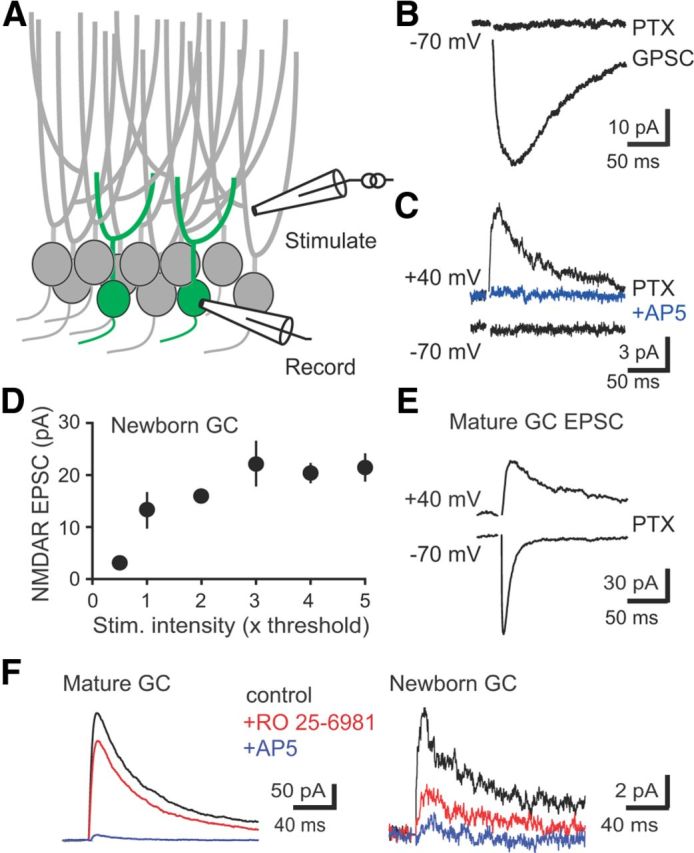
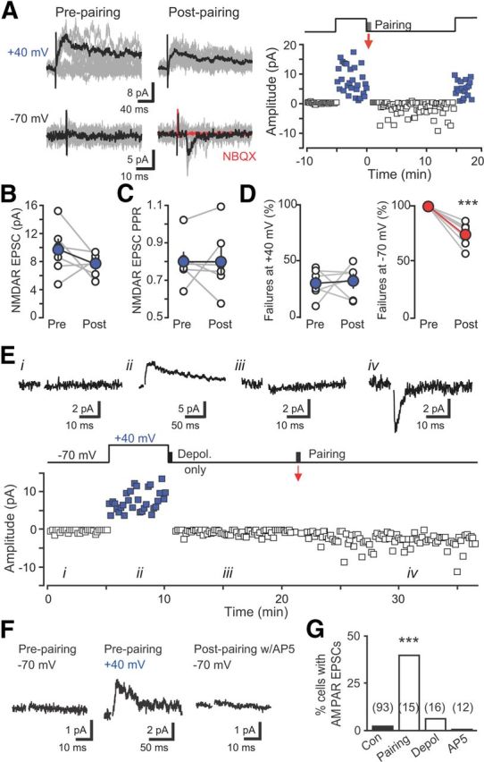
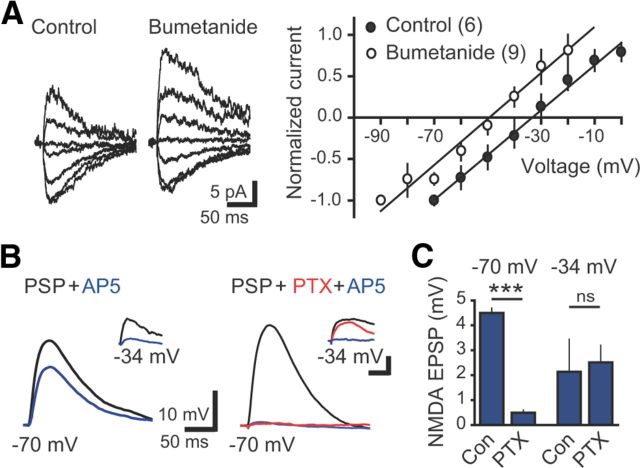
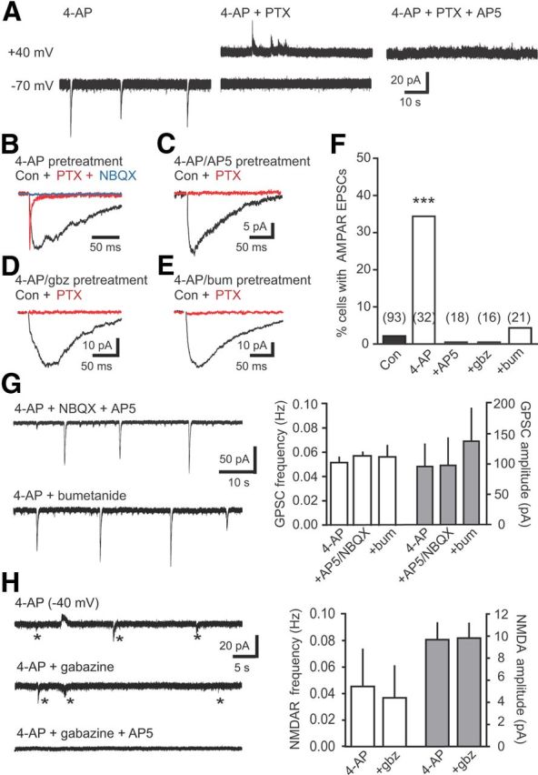
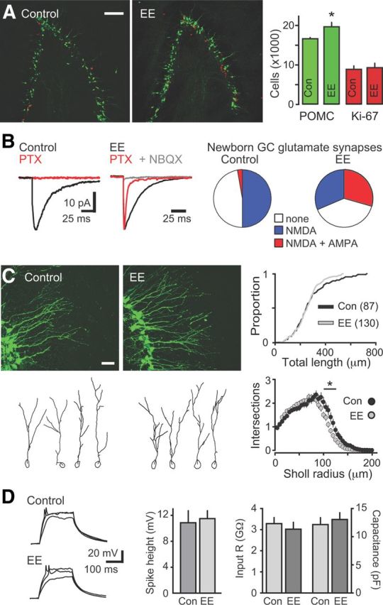
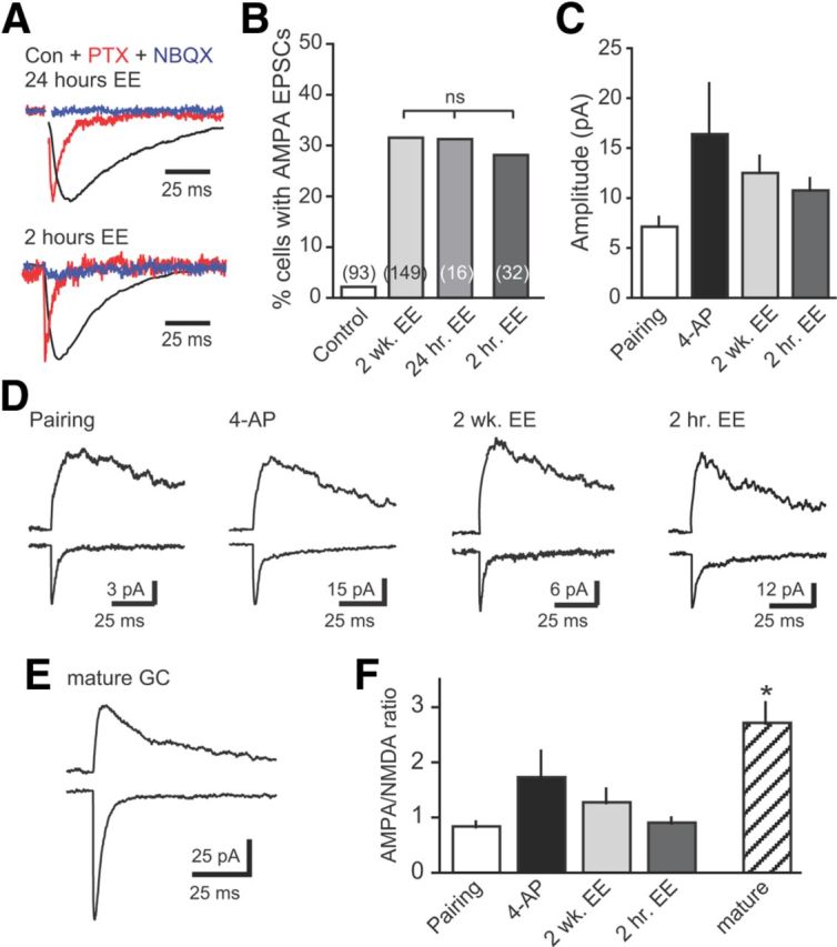
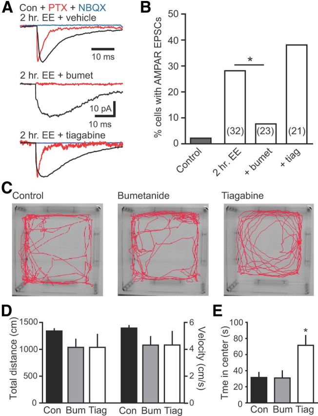
Neural activity enhances adult neurogenesis, enabling experience to influence the construction of new circuits. GABAA receptor-mediated depolarization of newborn neurons in the adult and developing brain promotes glutamatergic synaptic integration since chronic reduction of GABA depolarization impairs morphological maturation and formation of glutamatergic synapses. Here we demonstrate an acute role of GABA depolarization in glutamatergic synaptic integration. Using proopiomelanocortin enhanced-green fluorescent protein reporter mice, we identify a developmental stage when adult-generated neurons have glutamatergic synaptic transmission mediated solely by NMDA receptors (NMDARs), representing the initial silent synapses before AMPA receptor (AMPAR)-mediated functional transmission. We show that pairing synaptic stimulation with postsynaptic depolarization results in synapse unsilencing that requires NMDAR activation. GABA synaptic depolarization enables activation of NMDARs in the absence of AMPAR-mediated transmission and is required for synapse unsilencing induced by synaptic activity in vitro as well as a brief exposure to an enriched environment in vivo. The rapid appearance of AMPAR-mediated EPSCs and the lack of maturational changes show that GABA depolarization acutely allows NMDAR activation required for initial synapse unsilencing. Together, these results also reveal that adult-generated neurons in a critical period for survival use GABA signaling to rapidly initiate functional glutamate-mediated transmission in response to experience.


Figures

Figure 1.
Figure 1.
Newborn GCs have silent NMDAR-only synapses. A, Diagram of recording configuration. B, Synaptic stimulation near newborn GC dendrites evoked GPSCs blocked by PTX (100 μm; −70 mV holding potential). C, A typical silent synapse in PTX, with an NMDAR EPSC at +40 mV but no AMPAR EPSC at −70 mV. NMDAR EPSCs were blocked by AP5. D, The amplitude of NMDAR EPSCs in newborn GCs did not increase substantially with increased stimulation intensity (n = 5 cells with silent synapses), suggesting that very few synapses were present. Stimulus intensity was normalized to the intensity at which the NMDAR EPSC was just detectable (1× threshold). E, Similar stimulation intensities evoked dual AMPAR/NMDAR EPSCs in all neighboring mature GCs (n = 15). F, Differential block of NMDAR EPSCs in mature and immature GCs by Ro 25-6981. NMDAR EPSCs in mature GCs were recorded in NBQX.
Figure 2.
Figure 2.
Initial synapse unsilencing requires NMDAR activation. A, Left, EPSCs before and after pairing reveal rapid incorporation of AMPARs. Ten traces overlaid (gray) with averages (black). Right, Plot of EPSC amplitude versus time at −70 mV (black) and +40 mV (blue). Pairing protocol (red arrow) consisted of postsynaptic depolarization to 0 mV for 300 ms with presynaptic stimulation repeated at 1 Hz for 30 s. Baseline noise was subtracted. B, C, Neither the NMDAR EPSC amplitude (B) nor paired-pulse ratio (C) was altered by pairing. D, Failures of the NMDAR EPSC were unaffected by the pairing protocol (measured as the number of successes/total number of stimulations), whereas failures of the AMPAR EPSC were reduced. ***p < 0.001, paired t tests. E, Example EPSCs from the indicated times (above) and amplitude time course (below) showing that depolarization alone did not induce AMPA EPSCs that subsequently were induced by pairing depolarization with presynaptic stimulation. Artifacts blanked for clarity. F, Synapse unsilencing by the pairing protocol was blocked by AP5 (50 μm). G, Summary graph showing the percentage of newborn GCs with AMPAR EPSCs in each condition. The number of cells tested is shown in parentheses. ***p < 0.001, χ2 test compared with control. Con, Control; Depol, Depolarization.
Figure 3.
Figure 3.
Synaptic GABAergic depolarization allows activation of NMDARs. A, Left, Examples of GABAergic PSCs at holding potentials between −70 and 0 mV in gramicidin perforated patch recordings from newborn GCs in control and bumetanide-treated slices. NBQX (5 μm) and AP5 (50 μm) were used to isolate GPSCs. Right, Bumetanide (10 μm) hyperpolarized the reversal potential of GPSCs. B, Left, PSPs measured at −70 mV were partially blocked by AP5, indicating an NMDAR-mediated component. Right, PTX blocked PSP at −70 mV, although NMDAR EPSPs measured near EGABA (approximately −34 mV) were unaffected by PTX (insets; calibration: 2 mV, 100 ms). C, Right, Quantification of the AP5-sensitive component of PSPs recorded in control ACSF (n = 4) and PTX (n = 5) at each membrane potential. ***p < 0.001; ns, not significant; t test. Con, Control.
Figure 4.
Figure 4.
GABA depolarization is required for initial synapse unsilencing by synaptic activity. A, 4-AP (100 μm) induced rhythmic GABAR activation blocked by PTX in newborn GCs held at −70 mV. Subsequent depolarization to +40 mV revealed NMDAR EPSCs blocked by AP5 (50 μm). B, Example synaptic currents in a newborn GC after washout of 4-AP after 2 h incubation. Addition of PTX revealed an AMPAR EPSC (red; normalized to peak of GPSC) that was blocked by NBQX (10 μm; blue). C–E, Examples of synaptic currents in newborn GCs after incubation in 4-AP plus AP5 (50 μm; C), 4-AP plus gabazine (5 μm; D), or 4-AP plus bumetanide (10 μm; E). F, Summary of the percentage of newborn GCs with AMPAR EPSCs in control ASCF (black) or after 4-AP incubation with inclusion of the indicated antagonists (white). The number of cells tested is shown in parentheses. ***p < 0.001, χ2 test, compared with control. G, Left, Examples of rhythmic GABA release induced by 4-AP in the presence of blockers that prevent AMPAR incorporation. Right, The NKCC1 antagonist bumetanide (10 μm; n = 4) and the NMDAR and AMPAR antagonists AP5/NBQX (50 and 5 μm, respectively; n = 4) did not alter the frequency and amplitude of rhythmic GABAR PSCs (ANOVA). H, Left, Example of NMDAR-mediated EPSCs (asterisks) in a newborn GC held near the GABA reversal potential (small GPSCs are outward) in the indicated conditions. Right, Gabazine did not alter the frequency or amplitude of NMDAR EPSCs (n = 4, paired t test). bum, Bumetanide; Con, control; gbz, gabazine.
Figure 5.
Figure 5.
EE enhances newborn GC survival and promotes and initial synapse unsilencing in vivo. A, Left, POMC–GFP+ (green) and Ki67+ (red) expression identify newborn GCs and proliferative cells, respectively, in the DG of mice housed in standard environment (control; Con) and EE. Scale bar, 100 μm. Right, EE increased the number of GFP+ cells (n = 7 control and EE mice, p < 0.05, Mann–Whitney U test) without changing the number of Ki67+ cells (n = 3 control and EE mice, p = 0.78). B, PTX (red) blocked synaptic currents in newborn GCs from control mice (left), whereas NBQX-sensitive AMPAR EPSCs were present in newborn GCs from EE mice (middle). Right, EE increased the percentage of newborn GCs with functional AMPAR-containing synapses (p < 0.0001) and decreased the percentage of newborn GCs with no response (p < 0.05, χ2 test, n = 93 newborn GCs in control and 147 in EE). C, Left, Representative confocal images (top) and dendrite tracings (bottom) of newborn GCs from control and EE mice. Scale bar, 20 μm. Right top, The cumulative distribution of TDLs was not altered by EE, and there was no difference in the TDL (281 ± 15 vs 261 ± 9 μm), farthest extent of the dendrites (119 ± 4 vs 113 ± 2 μm), or number of nodes (5.8 ± 0.4 vs 5.5 ± 0.3; p > 0.05, unpaired t tests). Right bottom, There was a slight reduction in intersections at a distance of 95–120 μm from the soma in newborn GCs after EE (*p < 0.05). D, Left, Examples of current injections (40–60 pA) in newborn GCs from control and EE mice. Middle, EE did not alter the percentage of cells that fired action potentials (p = 0.12, χ2 test, n = 19 control and 25 EE) or the amplitude of spikes (0.76, unpaired t test). Right, The input resistance (p = 0.54) and capacitance (p = 0.59) were not different between newborn GCs in control and EE mice, indicating that newborn GCs were at the same developmental stage.
Figure 6.
Figure 6.
Short exposure to EE is sufficient for unsilencing in vivo. A, Examples of synaptic currents in newborn GCs after 24 or 2 h of EE, demonstrating that a short exposure to EE is sufficient for synapse unsilencing. EPSCs insensitive to PTX (red; normalized to peak of GPSC) were blocked by NBQX (5 μm; blue). B, Summary of the percentage of newborn GCs with AMPA EPSCs after exposure to EE for 2 weeks (data from Fig. 5), 24 h, or 2 h. C, The amplitude of AMPA EPSCs induced by pairing, 4-AP-driven synaptic activity, and EE was similar (p = 0.22, one-way ANOVA). D, Examples of AMPAR and NMDAR EPSCs in newborn GCs, measured at −70 mV and at +40 mV, respectively. E, Example of AMPAR and NMDAR EPSCs in a neighboring mature GC. F, The AMPA/NMDA ratio was similar in newborn GCs across all conditions (p = 0.16, ANOVA) but lower than in mature GCs (n = 15; *p < 0.01, t test of all newborn GCs compared with mature GCs). Con, Control.
Figure 7.
Figure 7.
GABAergic depolarization is necessary for synapse unsilencing by EE. A, Examples of synaptic currents in newborn GCs with vehicle, bumetanide (30 mg/kg, i.p.; middle) or tiagabine (10 mg/kg, i.p.) treatment before 2 h of EE. B, Summary of the percentage of newborn GCs with AMPAR EPSCs in each condition. Bumetanide reduced the percentage of newborn GCs with EE-induced AMPA EPSCs, whereas tiagabine had no significant effect. The number of cells tested is shown in parentheses. *p < 0.05, χ2 test. There was no difference in the percentage of newborn GCs with AMPAR EPSCs in the 2 h EE uninjected and vehicle-injected control groups (4 of 15 and 5 of 17, respectively; p = 1.0, χ2 test), so they were combined (n = 32). C, A separate group of mice were given vehicle (control; n = 7), bumetanide (30 mg/kg; n = 7), or tiagabine (10 mg/kg; n = 6) before exploring a novel open field for 4 min. D, E, Neither bumetanide nor tiagabine altered the total distance traveled and velocity (D; distance, p = 0.3; velocity, p = 0.3, ANOVA), but tiagabine increased the time spent in the center (E; p < 0.05). bumet/Bum, Bumetanide; Con, control; tiag/Tiag, tiagabine.

References

    1. Akerman CJ, Cline HT. Depolarizing GABAergic conductances regulate the balance of excitation to inhibition in the developing retinotectal circuit in vivo. J Neurosci. 2006;26:5117–5130. doi: 10.1523/JNEUROSCI.0319-06.2006. - DOI - PMC - PubMed
    1. Alme CB, Buzzetti RA, Marrone DF, Leutgeb JK, Chawla MK, Schaner MJ, Bohanick JD, Khoboko T, Leutgeb S, Moser EI, Moser MB, McNaughton BL, Barnes CA. Hippocampal granule cells opt for early retirement. Hippocampus. 2010;20:1109–1123. doi: 10.1002/hipo.20810. - DOI - PubMed
    1. Ambrogini P, Cuppini R, Lattanzi D, Ciuffoli S, Frontini A, Fanelli M. Synaptogenesis in adult-generated hippocampal granule cells is affected by behavioral experiences. Hippocampus. 2010;20:799–810. doi: 10.1002/hipo.20679. - DOI - PubMed
    1. Anderson ML, Sisti HM, Curlik DM, 2nd, Shors TJ. Associative learning increases adult neurogenesis during a critical period. Eur J Neurosci. 2011;33:175–181. doi: 10.1111/j.1460-9568.2010.07486.x. - DOI - PMC - PubMed
    1. Ben-Ari Y, Khazipov R, Leinekugel X, Caillard O, Gaiarsa JL. GABAA, NMDA and AMPA receptors: a developmentally regulated “menage a trois.”. Trends Neurosci. 1997;20:523–529. doi: 10.1016/S0166-2236(97)01147-8. - DOI - PubMed

Publication types