. 2013 Aug;24(2):288-97.
doi: 10.1007/s12640-013-9391-6. Epub 2013 Apr 11.

Evaluating the role of neuronal nitric oxide synthase-containing striatal interneurons in methamphetamine-induced dopamine neurotoxicity

Affiliations

Evaluating the role of neuronal nitric oxide synthase-containing striatal interneurons in methamphetamine-induced dopamine neurotoxicity

Ashley N Fricks-Gleason et al. Neurotox Res. 2013 Aug.

Abstract

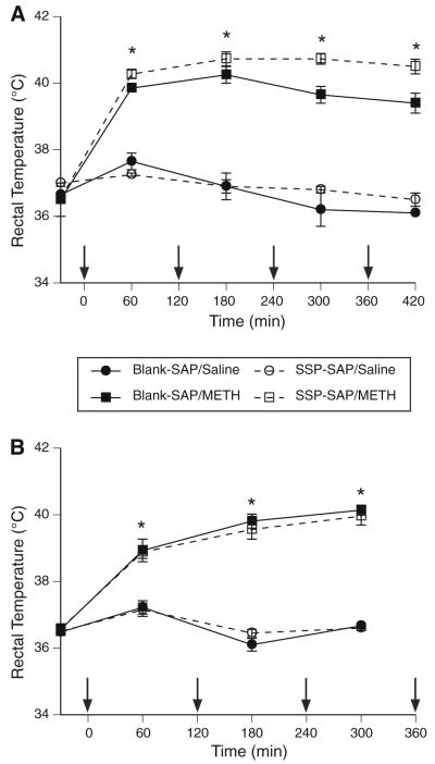
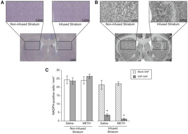
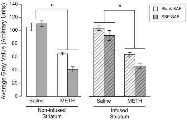
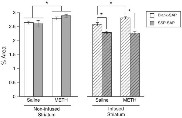
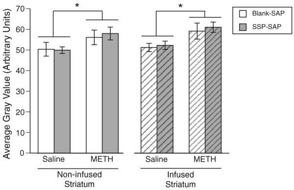
Production of nitric oxide (NO) has been implicated in methamphetamine (METH)-induced dopamine (DA) neurotoxicity. The source of this NO has not been clearly delineated, but recent evidence suggests that it arises from activation of neuronal nitric oxide synthase (nNOS), which is selectively expressed in a subpopulation of striatal interneurons. Our objective was to determine whether inhibiting activation of nNOS-containing interneurons in the striatum blocks METH-induced neurotoxicity. These interneurons selectively express the neurokinin-1 (NK-1) receptor, which is activated by substance P. One particular toxin, a conjugate of substance P to the ribosome-inactivating protein saporin (SSP-SAP), selectively destroys neurons expressing the NK-1 receptor. Thus, we examined the extent to which depletion of the nNOS-containing interneurons alters production of NO and attenuates METH-induced neurotoxicity. The SSP-SAP lesions resulted in significant loss of nNOS-containing interneurons throughout striatum. Surprisingly, this marked deletion did not confer resistance to METH-induced DA neurotoxicity, even in areas devoid of nNOS-positive cells. Furthermore, these lesions did not attenuate NO production, even in areas lacking nNOS. These data suggest that nNOS-containing interneurons either are not necessary for METH-induced DA neurotoxicity or produce NO that can diffuse extensively through striatal tissue and thereby still mediate neurotoxicity.


Figures

Fig. 1
Fig. 1
Body temperatures during METH administration. a Rectal temperatures over time for each of the four treatment groups sacrificed 7 days after the last injection: Blank-SAP/Saline (n = 2), Blank-SAP/METH (n = 2), SSP–SAP/Saline (n = 2), SSP–SAP/METH (n = 4). Rats were given intrastriatal infusions of Blank-SAP (control) or SSP–SAP (toxin) into one striatum followed by systemic administration of saline (4 × 1 ml/kg) or (±)-METH (4 × 7.5 mg/kg, s.c. at 2-hr intervals) 3 weeks later. * METH-treated rats significantly greater than saline-treated rats at each indicated time point, p < 0.05. b Rats were treated as described above for (a). Shown are rectal temperatures over time for each of the four treatment groups sacrificed 1 h after the last injection: Blank-SAP/Saline (n = 6), Blank-SAP/METH (n = 8), SSP–SAP/Saline (n = 6), SSP–SAP/METH (n = 9). * METH-treated rats significantly greater than saline-treated rats at each indicated time point, p < 0.05. Arrows indicate injection times
Fig. 2
Fig. 2
Representative photomicrographs from an animal that received SSP–SAP into the infused striatum followed by a systemic injection of saline three weeks later. a NADPH diaphorase histochemistry reveals a clear loss of nNOS-containing interneurons in the SSP–SAP-infused hemisphere. b NeuN IHC performed on an adjacent section confirms the specificity of the SSP–SAP-induced lesion. No gross loss of neurons was evident in the infused striatum other than mechanical damage from the cannulae. c Data are NADPH diaphorase histochemical staining in the striata of rats sacrificed 7 days after treatment with METH or saline (4 weeks after infusion of Blank-SAP or SSP–SAP into one striatum), and are represented as number of NADPH diaphorase-positive cell bodies per mm2 (mean ± SEM). * Significant main effect of pretreatment, p < 0.05
Fig. 3
Fig. 3
Effects of SSP–SAP infusions on METH-induced loss of DAT immunohistochemical staining in the striatum. Rats were treated as described in the legend to Fig. 1 and in the Methods. Data are DAT immunohistochemical staining in the striata of animals sacrificed 7 days after treatment with METH or saline (4 weeks after infusion of Blank-SAP or SSP–SAP into one striatum), and are represented as average gray values (mean ± SEM; arbitrary units). * Significant main effect of treatment, p < 0.05
Fig. 4
Fig. 4
Effects of SSP–SAP infusions on NADPH diaphorase histochemical staining in the striatum. Rats were treated as described in the legend to Fig. 1 and in the Methods. Data are NADPH diaphorase histochemical staining in the striata of animals sacrificed 1 h after treatment with METH or saline, and are represented as percent area of the imaged field with NADPH diaphorase histochemical signal (mean ± SEM). * Significant differences, p < 0.05
Fig. 5
Fig. 5
Effects of SSP–SAP infusions on nitrotyrosine (NT) immunohistochemical staining in the striatum. Rats were treated as described in the legend to Fig. 1 and in the Methods. Data are NT immunohistochemical staining in the striata of animals sacrificed 1 h after treatment with METH or saline, and are represented as average gray values (mean ± SEM; arbitrary units). * Significant main effect of treatment, p < 0.05

Similar articles

Cited by

References

    1. Abekawa T, Ohmori T, Koyama T. Effects of nitric oxide synthesis inhibition on methamphetamine-induced dopaminergic and serotonergic neurotoxicity in the rat brain. J Neural Transm. 1996;103(6):671–680. - PubMed
    1. Bergstrom BP, Garris PA. “Passive stabilization” of striatal extracellular dopamine across the lesion spectrum encompassing the presymptomatic phase of Parkinson’s disease: a voltammetric study in the 6-OHDA-lesioned rat. J Neurochem. 2003;87(5):1224–1236. - PubMed
    1. Bittner SE, Wagner GC, Aigner TG, Seiden LS. Effects of a high-dose treatment of methamphetamine on caudate dopamine and anorexia in rats. Pharmacol Biochem Behav. 1981;14(4):481–486. - PubMed
    1. Bowyer JF, Davies DL, Schmued L, Broening HW, Newport GD, Slikker W, Jr, Holson RR. Further studies of the role of hyperthermia in methamphetamine neurotoxicity. J Pharmacol Exp Ther. 1994;268(3):1571–1580. - PubMed
    1. Brown JM, Quinton MS, Yamamoto BK. Methamphetamine-induced inhibition of mitochondrial complex II: roles of glutamate and peroxynitrite. J Neurochem. 2005;95(2):429–436. doi:10.1111/j.1471-4159.2005.03379.x. - PubMed

Publication types