. 2013 Jun;87(12):6925-30.
doi: 10.1128/JVI.00283-13. Epub 2013 Apr 10.

Visualizing the beta interferon response in mice during infection with influenza A viruses expressing or lacking nonstructural protein 1

Affiliations

Visualizing the beta interferon response in mice during infection with influenza A viruses expressing or lacking nonstructural protein 1

Carsten Kallfass et al. J Virol. 2013 Jun.

Abstract

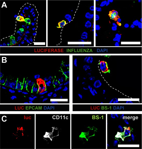
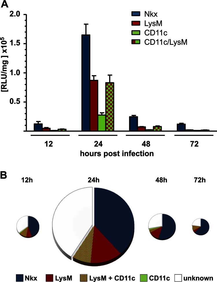
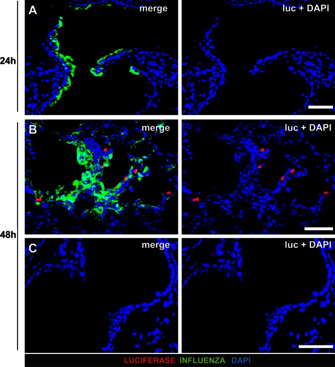
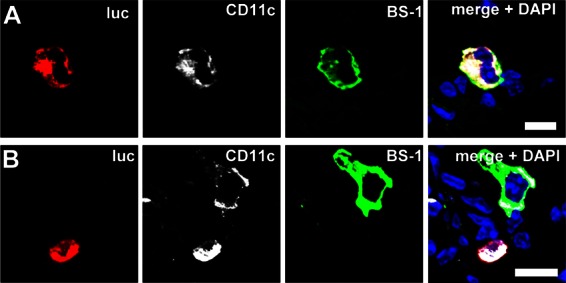
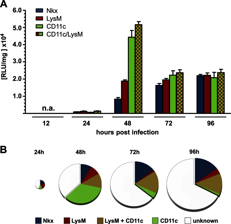
The innate host defense against influenza virus is largely dependent on the type I interferon (IFN) system. However, surprisingly little is known about the cellular source of IFN in the infected lung. To clarify this question, we employed a reporter mouse that contains the firefly luciferase gene in place of the IFN-β-coding region. IFN-β-producing cells were identified either by simultaneous immunostaining of lungs for luciferase and cellular markers or by generating conditional reporter mice that express luciferase exclusively in defined cell types. Two different strains of influenza A virus were employed that either do or do not code for nonstructural protein 1 (NS1), which strongly suppresses innate immune responses of infected cells. We found that epithelial cells and lung macrophages, which represent the prime host cells for influenza viruses, showed vigorous IFN-β responses which, however, were severely reduced and delayed if the infecting virus was able to produce NS1. Interestingly, CD11c(+) cell populations that were either expressing or lacking macrophage markers produced the bulk of IFN-β at 48 h after infection with wild-type influenza A virus. Our results demonstrate that the virus-encoded IFN-antagonistic factor NS1 disarms specifically epithelial cells and lung macrophages, which otherwise would serve as main mediators of the early response against infection by influenza virus.


Figures

Fig 1
Fig 1
Epithelial cells and macrophages are main producers of IFN-β in lungs of mice infected with SC35M-ΔNS1. Infected global reporter mice were sacrificed at 24 h postinfection, and lung slices were simultaneously stained for luciferase and either viral antigen (A) or cellular markers (B and C). (A) Luciferase-producing cells typically expressed viral antigen and were found in (left panel) or on top of (middle panel) the epithelium of bronchi and bronchioles. A few luciferase-positive cells were also found in the alveoli (right panel). (B) Luciferase-positive cells were either epithelial cells expressing EpCAM (left panel) or lectin BS-1-positive lung macrophages (right panel). (C) Luciferase-positive cells which expressed CD11c usually also reacted with BS-1, a lectin that can specifically bind to lung macrophages. Dashed lines indicate the apical surface of the lung epithelium. Staining of nuclei (blue) was achieved with DAPI. Size bars, 20 μm.
Fig 2
Fig 2
Conditional reporter mice confirm that epithelial cells and macrophages are the dominant IFN-β producers in mouse lungs during infection with SC35M-ΔNS1. Conditional reporter mice expressing Cre recombinase in epithelial cells (Nkx), macrophages/monocytes (LysM), CD11c+ cells (CD11c), or macrophages/monocytes and CD11c+ cells (LysM/CD11c) were infected with SC35M-ΔNS1. (A) Lungs were removed and tested for luciferase activity at the indicated time points. Values represent means ± standard errors of the means (SEM) for groups of animals (n ≥ 6) from each time point. (B) Luciferase values from infected conditional reporter mice expressing the luciferase gene exclusively in the indicated cell types were compared to luciferase values from infected “global” reporter mice. Values from “global” reporter mice were set to 100% and depicted as full-sized pies at each time point. The sizes of the pies are proportional to total luciferase activities at a particular time point postinfection.
Fig 3
Fig 3
IFN-β production in lungs of mice infected with wild-type SC35M. Lungs of infected “global” reporter mice were stained for luciferase and virus antigen. Counterstaining (blue) was done with DAPI. (A) No luciferase-positive cells were detectable at 24 h postinfection. (B and C) At 48 h postinfection, single dispersed luciferase-positive cells were detected in heavily infected (B) but not in virus antigen-free (C) lung areas. Note that luciferase-positive cells did not contain detectable levels of viral antigen. Size bars, 50 μm.
Fig 4
Fig 4
A fraction of CD11c+ cells producing IFN-β in lungs of mice infected with wild-type SC35M are macrophages. Lungs of virus-infected “global” reporter mice were harvested at 48 h postinfection and stained simultaneously with antibodies against luciferase (luc) and CD11c and with lectin BS-1. Luciferase-positive cells were positive for both CD11c and the lung macrophage marker BS-1 (A) or for CD11c but not BS-1 (B). Counterstaining (blue) was done with DAPI. Size bars, 10 μm.
Fig 5
Fig 5
Lung CD11c+ cells are the main producers of IFN-β at late stages of infection with wild-type SC35M. Conditional reporter mice selectively expressing luciferase in epithelial cells (Nkx), macrophages/monocytes (LysM), CD11c+ cells (CD11c), or macrophages/monocytes and CD11c+ cells (LysM/CD11c) were infected with wild-type SC35M, and luciferase activity in lungs was determined at the indicated time points. Values represent means ± SEM for groups of animals (n ≥ 6) from each time point. (B) Luciferase values from infected conditional mice expressing the reporter gene in the indicated cell types were compared to luciferase values from infected global reporter mice. Values from global reporter mice were set to 100% and depicted as full-sized pies at each time point. The sizes of the pies are proportional to total luciferase activity at a particular time point postinfection.

References

    1. Haller O, Kochs G, Weber F. 2006. The interferon response circuit: induction and suppression by pathogenic viruses. Virology 344:119–130 - PMC - PubMed
    1. Genin P, Vaccaro A, Civas A. 2009. The role of differential expression of human interferon-α genes in antiviral immunity. Cytokine Growth Factor Rev. 20:283–295 - PubMed
    1. Kallfass C, Ackerman A, Lienenklaus S, Weiss S, Heimrich B, Staeheli P. 2012. Visualizing production of Beta interferon by astrocytes and microglia in brain of la crosse virus-infected mice. J. Virol. 86:11223–11230 - PMC - PubMed
    1. Solodova E, Jablonska J, Weiss S, Lienenklaus S. 2011. Production of IFN-beta during Listeria monocytogenes infection is restricted to monocyte/macrophage lineage. PLoS One 6:e18543 doi:10.1371/journal.pone.0018543 - DOI - PMC - PubMed
    1. Erlandsson L, Blumenthal R, Eloranta ML, Engel H, Alm G, Weiss S, Leanderson T. 1998. Interferon-beta is required for interferon-alpha production in mouse fibroblasts. Curr. Biol. 8:223–226 - PubMed

Publication types

MeSH terms

LinkOut - more resources