A novel model for evaluating therapies targeting human tumor vasculature and human cancer stem-like cells
- PMID: 23576551
- PMCID: PMC3686886
- DOI: 10.1158/0008-5472.CAN-12-2845
A novel model for evaluating therapies targeting human tumor vasculature and human cancer stem-like cells
Abstract
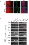
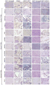
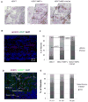
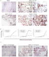
Human tumor vessels express tumor vascular markers (TVM), proteins that are not expressed in normal blood vessels. Antibodies targeting TVMs could act as potent therapeutics. Unfortunately, preclinical in vivo studies testing anti-human TVM therapies have been difficult to do due to a lack of in vivo models with confirmed expression of human TVMs. We therefore evaluated TVM expression in a human embryonic stem cell-derived teratoma (hESCT) tumor model previously shown to have human vessels. We now report that in the presence of tumor cells, hESCT tumor vessels express human TVMs. The addition of mouse embryonic fibroblasts and human tumor endothelial cells significantly increases the number of human tumor vessels. TVM induction is mostly tumor-type-specific with ovarian cancer cells inducing primarily ovarian TVMs, whereas breast cancer cells induce breast cancer specific TVMs. We show the use of this model to test an anti-human specific TVM immunotherapeutics; anti-human Thy1 TVM immunotherapy results in central tumor necrosis and a three-fold reduction in human tumor vascular density. Finally, we tested the ability of the hESCT model, with human tumor vascular niche, to enhance the engraftment rate of primary human ovarian cancer stem-like cells (CSC). ALDH(+) CSC from patients (n = 6) engrafted in hESCT within 4 to 12 weeks whereas none engrafted in the flank. ALDH(-) ovarian cancer cells showed no engraftment in the hESCT or flank (n = 3). Thus, this model represents a useful tool to test anti-human TVM therapy and evaluate in vivo human CSC tumor biology.
©2013 AACR.
Conflict of interest statement
Figures


Similar articles
-
Targeting tissue factor as a novel therapeutic oncotarget for eradication of cancer stem cells isolated from tumor cell lines, tumor xenografts and patients of breast, lung and ovarian cancer.Oncotarget. 2017 Jan 3;8(1):1481-1494. doi: 10.18632/oncotarget.13644. Oncotarget. 2017. PMID: 27903969 Free PMC article.
-
A novel experimental platform for investigating cancer growth and anti-cancer therapy in a human tissue microenvironment derived from human embryonic stem cells.Methods Mol Biol. 2006;331:329-46. doi: 10.1385/1-59745-046-4:329. Methods Mol Biol. 2006. PMID: 16881525
-
Ovarian cancer stem cells express ROR1, which can be targeted for anti-cancer-stem-cell therapy.Proc Natl Acad Sci U S A. 2014 Dec 2;111(48):17266-71. doi: 10.1073/pnas.1419599111. Epub 2014 Nov 19. Proc Natl Acad Sci U S A. 2014. PMID: 25411317 Free PMC article.
-
Florid vascular proliferation in mature cystic teratoma of the ovary: case report and review of the literature.Tumori. 2009 Jan-Feb;95(1):104-7. doi: 10.1177/030089160909500119. Tumori. 2009. PMID: 19366067 Review.
-
Role of the microenvironment in ovarian cancer stem cell maintenance.Biomed Res Int. 2013;2013:630782. doi: 10.1155/2013/630782. Epub 2012 Dec 24. Biomed Res Int. 2013. PMID: 23484135 Free PMC article. Review.
Cited by
-
Niche-dependent gene expression profile of intratumoral heterogeneous ovarian cancer stem cell populations.PLoS One. 2013 Dec 17;8(12):e83651. doi: 10.1371/journal.pone.0083651. eCollection 2013. PLoS One. 2013. PMID: 24358304 Free PMC article.
-
Streptavidin-Saporin: Converting Biotinylated Materials into Targeted Toxins.Toxins (Basel). 2023 Feb 27;15(3):181. doi: 10.3390/toxins15030181. Toxins (Basel). 2023. PMID: 36977072 Free PMC article. Review.
-
Hedgehog signaling underlying tendon and enthesis development and pathology.Matrix Biol. 2022 Jan;105:87-103. doi: 10.1016/j.matbio.2021.12.001. Epub 2021 Dec 24. Matrix Biol. 2022. PMID: 34954379 Free PMC article. Review.
-
Therapeutic Impact of Nanoparticle Therapy Targeting Tumor-Associated Macrophages.Mol Cancer Ther. 2018 Jan;17(1):96-106. doi: 10.1158/1535-7163.MCT-17-0688. Epub 2017 Nov 13. Mol Cancer Ther. 2018. PMID: 29133618 Free PMC article.
-
Targeting the Sonic Hedgehog Signaling Pathway: Review of Smoothened and GLI Inhibitors.Cancers (Basel). 2016 Feb 15;8(2):22. doi: 10.3390/cancers8020022. Cancers (Basel). 2016. PMID: 26891329 Free PMC article. Review.
References
-
- St Croix B, Rago C, Velculescu V, Traverso G, Romans KE, Montgomery E, et al. Genes expressed in human tumor endothelium. Science. 2000;289:1197–202. - PubMed
-
- Pasqualini R, Moeller BJ, Arap W. Leveraging molecular heterogeneity of the vascular endothelium for targeted drug delivery and imaging. Semin Thromb Hemost. 2010;36:343–51. - PubMed
-
- Allinen M, Beroukhim R, Cai L, Brennan C, Lahti-Domenici J, Huang H, et al. Molecular characterization of the tumor microenvironment in breast cancer.[see comment] Cancer Cell. 2004;6:17–32. - PubMed
Publication types
MeSH terms
Substances
Grants and funding
LinkOut - more resources
Full Text Sources
Other Literature Sources
Medical
Molecular Biology Databases
Miscellaneous

