. 2013 Jun 1;73(11):3381-92.
doi: 10.1158/0008-5472.CAN-12-4047. Epub 2013 Apr 10.

Hedgehog signaling alters reliance on EGF receptor signaling and mediates anti-EGFR therapeutic resistance in head and neck cancer

Affiliations

Hedgehog signaling alters reliance on EGF receptor signaling and mediates anti-EGFR therapeutic resistance in head and neck cancer

Stephen B Keysar et al. Cancer Res. .

Abstract

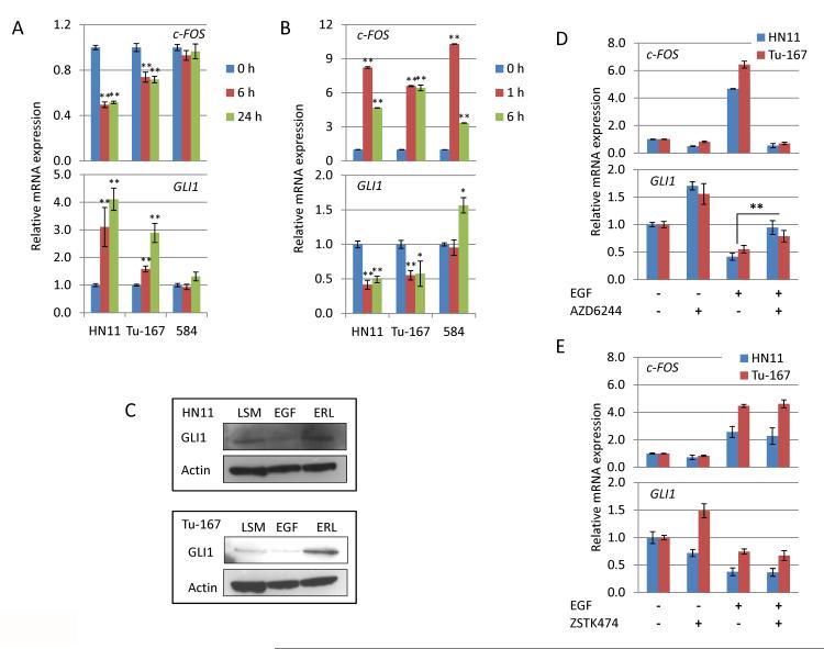
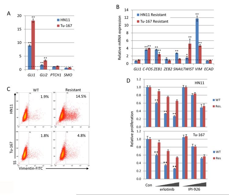
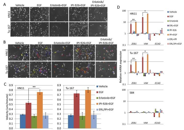
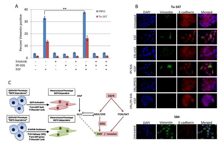
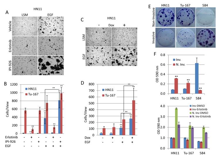
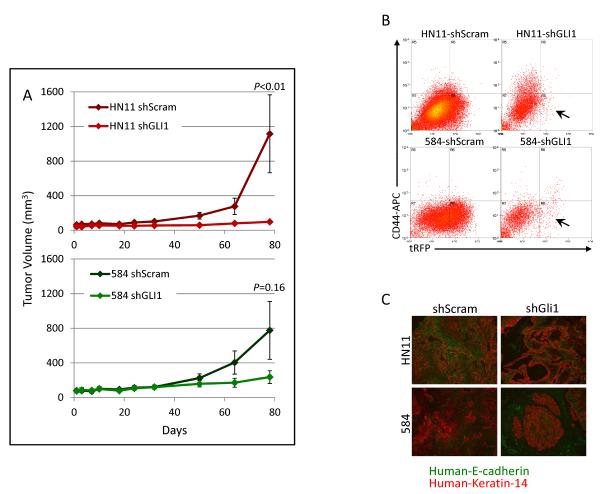
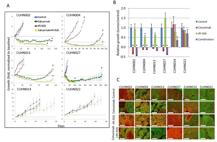
The EGF receptor (EGFR)-directed monoclonal antibody cetuximab is the only targeted therapy approved for the treatment of squamous cell carcinoma of the head and neck (HNSCC) but is only effective in a minority of patients. Epithelial-to-mesenchymal transition (EMT) has been implicated as a drug resistance mechanism in multiple cancers, and the EGFR and Hedgehog pathways (HhP) are relevant to this process, but the interplay between the two pathways has not been defined in HNSCC. Here, we show that HNSCC cells that were naturally sensitive to EGFR inhibition over time developed increased expression of the HhP transcription factor GLI1 as they became resistant after long-term EGFR inhibitor exposure. This robustly correlated with an increase in vimentin expression. Conversely, the HhP negatively regulated an EGFR-dependent, EMT-like state in HNSCC cells, and pharmacologic or genetic inhibition of HhP signaling pushed cells further into an EGFR-dependent phenotype, increasing expression of ZEB1 and VIM. In vivo treatment with cetuximab resulted in tumor shrinkage in four of six HNSCC patient-derived xenografts; however, they eventually regrew. Cetuximab in combination with the HhP inhibitor IPI-926 eliminated tumors in two cases and significantly delayed regrowth in the other two cases. Expression of EMT genes TWIST and ZEB2 was increased in sensitive xenografts, suggesting a possible resistant mesenchymal population. In summary, we report that EGFR-dependent HNSCC cells can undergo both EGFR-dependent and -independent EMT and HhP signaling is a regulator in both processes. Cetuximab plus IPI-926 forces tumor cells into an EGFR-dependent state, delaying or completely blocking tumor recurrence.


Figures

Figure 1
Figure 1. Inhibition of EGFR signaling increases expression of HhP gene in EGFR dependent cells
A. 1μM erlotinib suppresses c-FOS expression in both HN11 and Tu-167 cells, but not 584 cells, at 6 and 24h. Expression of GLI1 is significantly increased in both HN11 and Tu-167 cells but is unchanged in 584 cells. B. EGFR activation by EGF (100ng/ml) induces c-FOS expression in all cell lines within 1h and significantly suppresses GLI1 levels in HN11 and Tu-167 cells at both 1 and 6h. However, GLI1 levels are increased in 584 cells at 6h after EGF treatment. C. 96h treatment with EGF and erlotinib suppresses and increase GLI1 levels respectively in both HN11 and Tu-167 cells. D and E. Suppression of GLI1 by EGFR activation occurs through the MEK/ERK pathway as the MEK inhibitor AZD6244 blocks suppression of Gli1 by EGF while the PI3K inhibitor ZSTK474 does not. *P<0.05, **P<0.01. All significance was calculated to control unless indicated by a bar between experimental groups.
Figure 2
Figure 2. Chronic erlotinib treatment generates resistant cells with a mesenchymal gene expression pattern
A. Expression of GLI transcription factors are significantly increased in EGFR dependent cells chronically treated with 10μM erlotinib. B. Erlotinib resistant cells highly express pro-EMT genes after removal from selection media. C. Percentage of Vim-positive cells is significantly increased in erlotinib resistant cell lines, measured by flow cytometry D. Erlotinib resistant cells are not sensitized to HhP inhibition by IPI-926. Cells were treated with erlotinib (1,5,10μM) or IPI-926 (1,5,10μM) and proliferation was measured by the SRB assay. Sensitivity to erlotonib decreased in resistant cells while sensitivity to IPI-926 was unchanged. *P<0.05, **P<0.01.
Figure 3
Figure 3. Inhibition of the hedgehog pathway may augment EGFR driven EMT-like state in EGFR dependent cell lines
A. EGFR signaling activation by the EGF ligand (100ng/ml) generates an EMT-like phenotype in HN11 and Tu-167 cells that is completely blocked by erlotinib (1μM) but not IPI-926 (1μM). B and C. EGF increases cellular motility in EGFR dependent cells which was blocked by erlotinib but augmented by IPI-926. D. EGF increased expression of pro-EMT genes ZEB1 and VIM as well as suppressed E-cad generating an EMT-like gene expression profile in HN11 and Tu-167 cells but not 584 cells. However, E-CAD expression was suppressed by erlotinib treatment in 584 cells. *P<0.05, **P<0.01.
Figure 4
Figure 4. EGFR driven EMT-like state is associated with Vimentin-positive cells
A. EGF increased the percentage of Vim-positive cells in HN11 and Tu-167 cell lines measured by flow cytometry. Erlotinib blocked this effect while IPI-926 significantly increased the positive population in HN11 cells. B. EGF increased the number of Vim-positive cells and induced cellular dissociation and slight decreases in E-cad. 584 cells are all positive for Vim and express very low levels of E-cad. C. Schemata depicting (Left) EGFR-dependent and -independent EMT and (Right) the relevant pathways downstream of EGFR. *P<0.05, **P<0.01.
Figure 5
Figure 5. EGF drives invasion in EGFR dependent cells and is augmented by inhibition of the HhP
A and B. Erlotinib blocks EGF-induced invasion while IPI-926 significantly increases invasion by HN11 cells. C and D. Expression of shRNA against GLI1 increased EGF-induced invasion in HN11 and Tu-167 cells. E and F. Invasive cells are less proliferative than non-invasive cells from the EGFR dependent cell lines HN11 and Tu-167. The opposite is true for the mesenchymal 584 cells. *P<0.05, **P<0.01.
Figure 6
Figure 6. Silencing of GLI1 inhibits tumor formation in nude mice
A. Tumor formation of HN11 and 584 cells expressing shRNA against GLI1 was suppressed when compared to scrambled controls. B. shRNA expressing cells simultaneously express turboRFP, which was nearly completely lost in shGli1 expressing tumors compared to scrambled controls. C. shGli1 expressing tumors contained fewer tumors cells and higher levels of mouse stroma when compared to tumors expressing a scrambled sequence.
Figure 7
Figure 7. Combination treatment of human HNSCC xenografts with cetuximab and IPI-926 blocks tumor re-growth
A. Growth kinetics of 6 HNSCC xenograft cases treated with cetuximab, IPI-926 and cetuximab+IPI-926. B. Tumor/Control values for treated cases. C. Staining of E-cadherin (green) and Keratin-14 (red) in HNSCC xenografts after treatment. Treatment with IPI-926 decreased E-cadherin, pushing cells into an EGFR-driven EMT-like state. Scaling bars represent 50μm at 40×. *P<0.05.

Similar articles

Cited by

References

    1. Siegel R, Naishadham D, Jemal A. Cancer statistics, 2012. CA Cancer J Clin. 2012;62:10–29. - PubMed
    1. Bonner JA, Harari PM, Giralt J, Azarnia N, Shin DM, Cohen RB, et al. Radiotherapy plus cetuximab for squamous-cell carcinoma of the head and neck. The New England journal of medicine. 2006;354:567–78. - PubMed
    1. Vermorken JB, Mesia R, Rivera F, Remenar E, Kawecki A, Rottey S, et al. Platinum-based chemotherapy plus cetuximab in head and neck cancer. The New England journal of medicine. 2008;359:1116–27. - PubMed
    1. Wheeler DL, Huang S, Kruser TJ, Nechrebecki MM, Armstrong EA, Benavente S, et al. Mechanisms of acquired resistance to cetuximab: role of HER (ErbB) family members. Oncogene. 2008;27:3944–56. - PMC - PubMed
    1. Benavente S, Huang S, Armstrong EA, Chi A, Hsu KT, Wheeler DL, et al. Establishment and characterization of a model of acquired resistance to epidermal growth factor receptor targeting agents in human cancer cells. Clin Cancer Res. 2009;15:1585–92. - PMC - PubMed

Publication types

MeSH terms