. 2013 Jun 15;73(12):3692-703.
doi: 10.1158/0008-5472.CAN-12-2160. Epub 2013 Apr 10.

Inhibition of c-Met reduces lymphatic metastasis in RIP-Tag2 transgenic mice

Affiliations

Inhibition of c-Met reduces lymphatic metastasis in RIP-Tag2 transgenic mice

Barbara Sennino et al. Cancer Res. .

Abstract

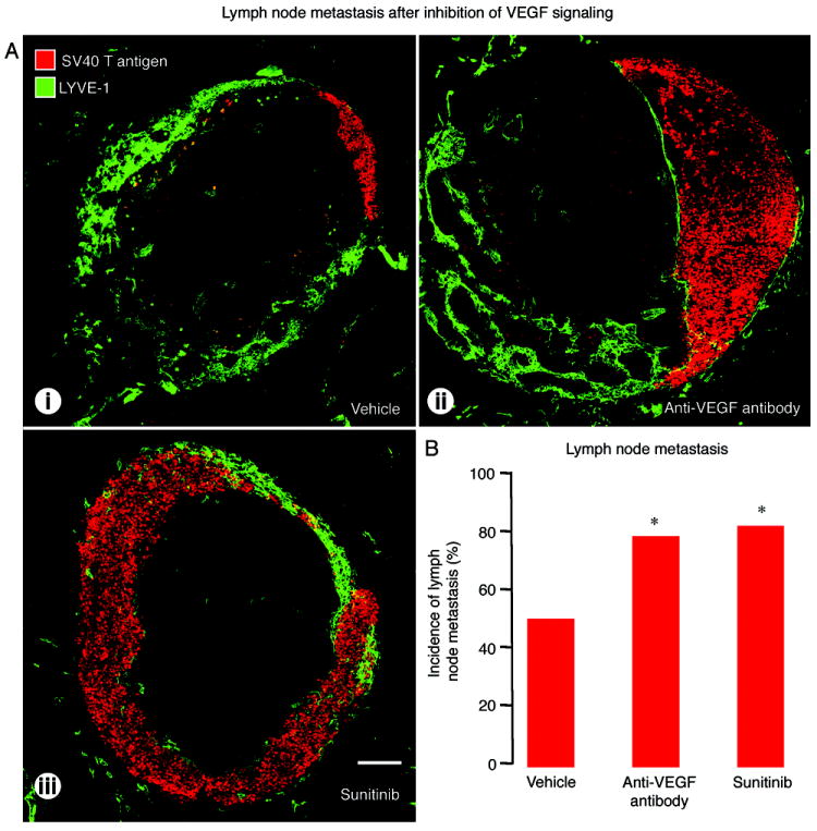
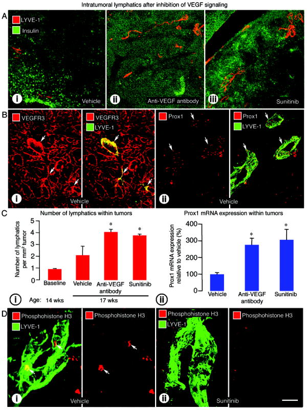
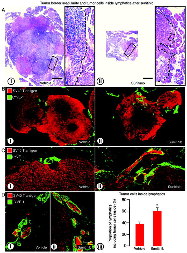
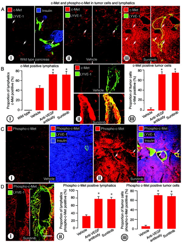
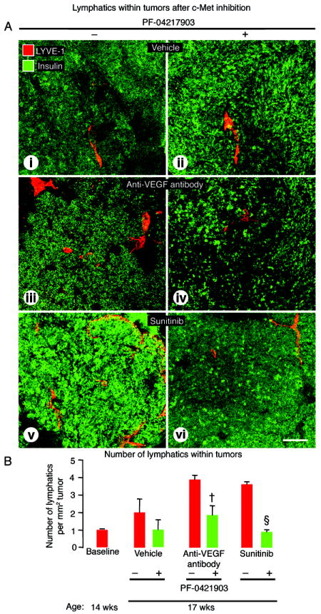
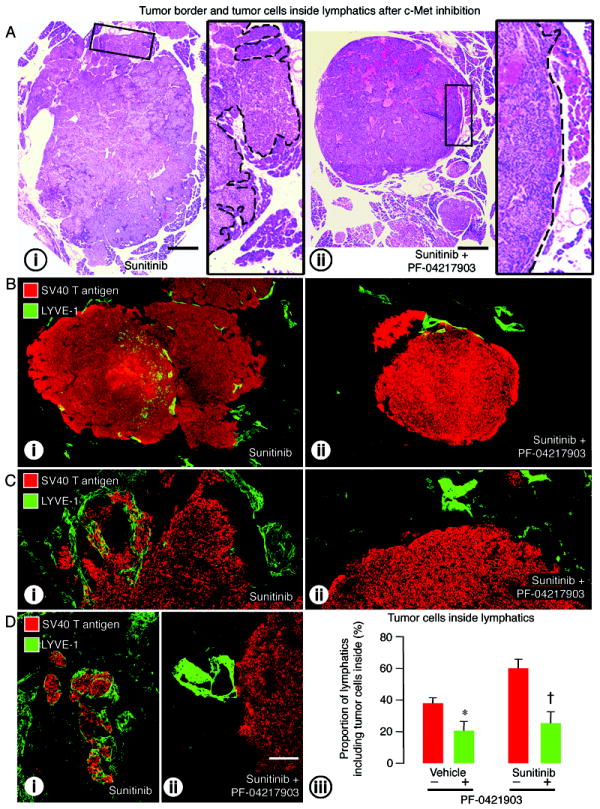
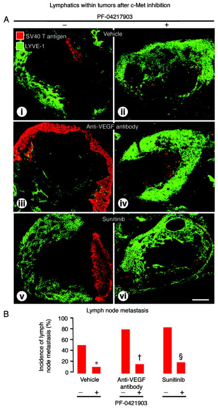
Inhibition of VEGF signaling can promote lymph node metastasis in preclinical models, but the mechanism is not fully understood, and successful methods of prevention have not been found. Signaling of hepatocyte growth factor (HGF) and its receptor c-Met can promote the growth of lymphatics and metastasis of some tumors. We sought to explore the contributions of c-Met signaling to lymph node metastasis after inhibition of VEGF signaling. In particular, we examined whether c-Met is upregulated in lymphatics in or near pancreatic neuroendocrine tumors in RIP-Tag2 transgenic mice and whether lymph node metastasis can be reduced by concurrent inhibition of VEGF and c-Met signaling. Inhibition of VEGF signaling by anti-VEGF antibody or sunitinib in mice from the age of 14 to 17 weeks was accompanied by more intratumoral lymphatics, more tumor cells inside lymphatics, and more lymph node metastases. Under these conditions, lymphatic endothelial cells, like tumor cells, had strong immunoreactivity for c-Met and phospho-c-Met. c-Met blockade by the selective inhibitor, PF-04217903, significantly reduced metastasis to local lymph nodes. Together, these results indicate that inhibition of VEGF signaling in RIP-Tag2 mice upregulates c-Met expression in lymphatic endothelial cells, increases the number of intratumoral lymphatics and number of tumor cells within lymphatics, and promotes metastasis to local lymph nodes. Prevention of lymph node metastasis by PF-04217903 in this setting implicates c-Met signaling in tumor cell spread to lymph nodes.


Conflict of interest statement

Disclosure of Potential Conflicts of Interest

J. Christensen: employee of Pfizer Global Research and Development. D.M. McDonald: research funding from a grant to University of California- San Francisco from Pfizer Global Research and Development. The other authors had no potential conflicts of interest to disclose.

Figures

Figure 1
Figure 1
Effect of VEGF signaling inhibition on metastases in pancreatic lymph nodes of RIP-Tag2 mice treated from age 14 to 17 weeks. A: Confocal micrographs of lymph nodes (LYVE-1, green) with metastases (SV40 T-antigen, red) after treatment with (i) vehicle, (ii) anti-VEGF antibody, or (iii) sunitinib. B: Proportion of lymph nodes with metastases showing significantly higher incidence of metastasis after anti-VEGF antibody or sunitinib. *P<0.05 compared to vehicle. Scale bar in A-iii applies to all images: 200μm.
Figure 2
Figure 2
Effect of VEGF signaling inhibition on lymphatics inside RIP-Tag2 tumors. A: Confocal micrographs of intratumoral lymphatics (LYVE-1, red) in tumors (insulin, green) after treatment with (i) vehicle, (ii) anti-VEGF antibody, or (iii) sunitinib from age 14 to 17 weeks. B: (i) VEGFR-3 immunoreactivity (red) in abundant tumor blood vessels and a few lymphatic vessels (LYVE-1, green, arrows); (ii) Prox1 immunoreactivity (red) in lymphatic vessels (LYVE-1, green, arrows) in vehicle-treated RIP-Tag2 tumors. C: (i) Measurements showing significantly higher density of lymphatics inside tumors treated with anti-VEGF antibody or sunitinib from age 14 to 17 weeks; (ii) qRT-PCR measurements showing significantly higher Prox1 mRNA expression in tumors treated with anti-VEGF antibody or sunitinib from age 14 to 15 weeks. D: Confocal images illustrating the presence of proliferating cells (phosphohistone H3, red, arrows) in a lymphatic vessel (LYVE-1, green) after vehicle (i) but not after sunitinib (ii). The colocalization of phosphohistone H3 stained nuclei and LYVE-1 stained lymphatic cells was confirmed by examination of individual optical slices. *P<0.05 compared to vehicle. Scale bar in D-ii applies to all images: 200μm for A; 50μm for B; 7μm for D.
Figure 3
Figure 3
Effect of VEGF signaling inhibition on tumor border irregularity and tumor cells inside lymphatics in RIP-Tag2 mice treated from age 14 to 17 weeks. A: Tumor sections stained with hematoxylin and eosin (H&E) comparing tumor size and tumor edges after treatment with (i) vehicle or (ii) sunitinib. Composite images of tumor sections (B) and confocal micrographs (C) showing the irregularity of tumor border (SV40 T-antigen, red) and tumor cells inside lymphatics (LYVE-1, green) after treatment with (i) vehicle or (ii) sunitinib. D: Tumor cells inside lymphatics after (i) vehicle or (ii) sunitinib. (iii) Measurements showing significantly higher proportion of lymphatics with tumor cells in the lumen after sunitinib. *P<0.05 compared to vehicle. Scale bar in A applies to Ai and Aii: 1.3mm for the large images and 200μm for inserts. Scale bar in D-ii applies to B-D: 800μm for B; 120μm for C; 200μm for D.
Figure 4
Figure 4
c-Met and phospho-c-Met immunoreactivities in tumor cells and lymphatics of RIP-Tag2 mice treated from age 14 to 17 weeks. A: (i) Wild-type pancreas (normal islets, insulin, blue) with lymphatic vessels (LYVE-1, green, arrows) lacking c-Met immunoreactivity (red). ii-iii: c-Met (red) in intratumoral lymphatics (LYVE-1, green, arrows) in tumors treated with (ii) vehicle or (iii) sunitinib. B: (i) Measurements showing significantly higher proportion of lymphatic vessels with c-Met immunoreactivity after anti-VEGF antibody or sunitinib. (ii) High magnification confocal images showing the difference in intensity of c-Met staining in lymphatics after vehicle and sunitinib treatment. (iii) Measurements showing higher proportion of tumor cells with c-Met immunoreactivity after anti-VEGF antibody or sunitinib. C: Comparison of phospho c-Met immunoreactivity (red) in tumor cells (insulin, blue) or intratumoral lymphatics (LYVE-1, green, arrows) after treatment with (i) vehicle or (ii) sunitinib. D: (i) Phospho c-Met staining (red) in the endothelium of an intratumoral lymphatic (LYVE-1, green) after treatment with sunitinib. (ii-iii) Bar graphs showing significantly higher proportion of phospho-c-Met immunoreactive (ii) lymphatic vessels and (iii) tumor cells after anti-VEGF antibody or sunitinib. *P<0.05 compared to vehicle. Scale bar in D-i applies to all images: 50μm for A and C; 8μm for C and D.
Figure 5
Figure 5
Effect of c-Met inhibition on lymphatics within RIP-Tag2 tumors after treatment from age 14 to 17 weeks. A: Confocal micrographs of intratumoral lymphatics (LYVE-1, red) in tumors (insulin, green) after treatment with (i-ii) vehicle, (iii-iv) anti-VEGF antibody, or (v-vi) sunitinib without (-) or with (+) c-Met inhibition by PF-04217903. B: Measurements showing significantly fewer lymphatics in tumors treated with PF-04217903. P<0.05 compared to anti-VEGF antibody (†) or sunitinib (§) without PF-04217903. Scale bar in A-vi applies to all images: 125μm.
Figure 6
Figure 6
Effect of c-Met inhibition on tumor border irregularity and lymphatics in RIP-Tag2 mice treated from age 14 to 17 weeks. A: Tumor sections stained with H&E comparing tumor size and border contour after treatment with (i) sunitinib alone or (ii) sunitinib plus PF-04217903. Composite images of tumor sections (B) and confocal micrographs (C) showing the tumor border (SV40 T-antigen, red) and lymphatic vessels (LYVE-1, green) after treatment with (i) sunitinib or (ii) sunitinib plus PF-04217903. D: Tumor cells inside lymphatics after (i) sunitinib alone (-) or (ii) sunitinib with PF-04217903 (+). (iii) Measurements showing significantly smaller proportion of lymphatics with tumor cells in the lumen after PF-04217903 administered with vehicle or sunitinib. P<0.05 compared to vehicle (*) or sunitinib (†) without PF-04217903. Scale bar in A applies to A-i and A-ii: 900μm for the large images and 200μm for inserts. Scale bar in D-ii applies to B-D: 500μm for B; 120μm for C; 200μm for D.
Figure 7
Figure 7
Effect of c-Met inhibition on pancreatic lymph node metastases in RIP-Tag2 mice treated from age 14 to 17 weeks. A: Confocal micrographs of lymph nodes (LYVE-1, green), some of which have metastases (SV40 T-antigen, red), after treatment with (i-ii) vehicle, (iii-iv) anti-VEGF antibody, or (v-vi) sunitinib without (-) or with (+) c-Met inhibition by PF-04217903. B: Measurements showing significantly smaller proportion of lymph nodes with metastases after PF-04217903 in all three groups. P<0.05 compared to vehicle (*), anti-VEGF antibody (†), or sunitinib (§) without PF-04217903. Scale bar in D-ii applies to all images: 220μm.

Similar articles

Cited by

References

    1. Cao Y. Opinion: emerging mechanisms of tumour lymphangiogenesis and lymphatic metastasis. Nat Rev Cancer. 2005;5:735–43. - PubMed
    1. Mandriota SJ, Jussila L, Jeltsch M, Compagni A, Baetens D, Prevo R, et al. Vascular endothelial growth factor-C-mediated lymphangiogenesis promotes tumour metastasis. EMBO J. 2001;20:672–82. - PMC - PubMed
    1. Skobe M, Hawighorst T, Jackson DG, Prevo R, Janes L, Velasco P, et al. Induction of tumor lymphangiogenesis by VEGF-C promotes breast cancer metastasis. Nat Med. 2001;7:192–8. - PubMed
    1. Stacker SA, Caesar C, Baldwin ME, Thornton GE, Williams RA, Prevo R, et al. VEGF-D promotes the metastatic spread of tumor cells via the lymphatics. Nat Med. 2001;7:186–91. - PubMed
    1. Casanovas O, Hicklin DJ, Bergers G, Hanahan D. Drug resistance by evasion of antiangiogenic targeting of VEGF signaling in late-stage pancreatic islet tumors. Cancer Cell. 2005;8:299–309. - PubMed

Publication types

MeSH terms