. 2013 Jun 1;73(11):3356-70.
doi: 10.1158/0008-5472.CAN-12-3904. Epub 2013 Apr 10.

Threshold levels of ABL tyrosine kinase inhibitors retained in chronic myeloid leukemia cells determine their commitment to apoptosis

Affiliations

Threshold levels of ABL tyrosine kinase inhibitors retained in chronic myeloid leukemia cells determine their commitment to apoptosis

Thomas O'Hare et al. Cancer Res. .

Abstract

The imatinib paradigm in chronic myelogenous leukemia (CML) established continuous BCR-ABL inhibition as a design principle for ABL tyrosine kinase inhibitors (TKI). However, clinical responses seen in patients treated with the ABL TKI dasatinib despite its much shorter plasma half-life and the apparent rapid restoration of BCR-ABL signaling activity following once-daily dosing suggested acute, potent inhibition of kinase activity may be sufficient to irrevocably commit CML cells to apoptosis. To determine the specific requirements for ABL TKI-induced CML cell death for a panel of clinically important ABL TKIs (imatinib, nilotinib, dasatinib, ponatinib, and DCC-2036), we interrogated response of CML cell lines and primary CML cells following acute drug exposure using intracellular fluorescence-activated cell sorting and immunoblot analyses of BCR-ABL signaling, apoptosis measurements, liquid chromatography/tandem mass spectrometry of intracellular drug levels, and biochemical TKI dissociation studies. Importantly, significant intracellular TKI stores were detected following drug washout, levels of which tracked with onset of apoptosis and incomplete return of BCR-ABL signaling, particularly pSTAT5, to baseline. Among TKIs tested, ponatinib showed the most robust capacity for apoptotic commitment showing sustained suppression of BCR-ABL signaling even at low intracellular levels following extensive washout, consistent with high-affinity binding and slow dissociation from ABL kinase. Together, our findings suggest commitment of CML cells to apoptosis requires protracted incomplete restoration of BCR-ABL signaling mediated by intracellular retention of TKIs above a quantifiable threshold. These studies refine our understanding of apoptotic commitment in CML cells and highlight parameters important to design of therapeutic kinase inhibitors for CML and other malignancies.


Figures

Figure 1
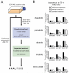
Figure 1. Commitment of BCR-ABL-positive cells to apoptosis following acute exposure to ABL TKIs varies by inhibitor, concentration, and extent of washout
(A) Schematic outlining general experimental washout protocol. Briefly, BCR-ABL-positive cells were incubated in complete media alone or in the presence of each of five clinically relevant ABL TKIs either continuously (72 h) or for a short exposure (2 h for cell lines, 4 h for primary cells). For acute exposure conditions, cells were subjected to our previously published 3-wash-step protocol (standard washout) followed by an additional 5 wash steps (expanded washout) and cultured in fresh complete media for the remainder of the 72 h experiment. (B) Levels of apoptosis in K562 cells following continuous or acute exposure to ABL kinase inhibitors. Cells were incubated in the presence of the indicated inhibitor concentrations for 2 h, and samples were collected just prior to washout and immediately following standard and expanded washout. Annexin V-positivity was measured at 72 h after the start of the experiment, and bars represent the mean of at least three independent experiments performed in triplicate ± S.E.M.
Figure 2
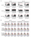
Figure 2. BCR-ABL signaling activity is not fully restored to baseline levels in treatment/washout conditions of dasatinib and ponatinib that commit cells to apoptosis
K562 cells were incubated alone or in the presence of 10 and 100 nM dasatinib or ponatinib for 2 h, subjected to standard and expanded washout, and collected at the indicated timepoint post washout for analysis by (A) immunoblot and (B) Phosflow FACS analyses. For immunoblot analyses, the phosphorylated and non-phosphorylated forms of CrkL were resolved by SDS-PAGE, blotted using a total CrkL antibody, and results are expressed as % pCrkL with the red, dashed line indicating the level of % pCrkL in untreated K562 cells. For Phosflow FACS analyses, cells were fixed, permeabilized, and stained using Alexa647-pCrkL and Alexa488-pSTAT5 conjugated antibodies. Results are displayed, for comparison purposes, as the overlaid signal peak traces of isotype control, untreated cells, and each indicated timepoint post washout. Vertical, black dashed lines highlight the peak signal in untreated K562 cells for reference.
Figure 3
Figure 3. Extent of restoration of BCR-ABL signaling activity following washout of less potent, more selective ABL TKIs tracks with conditions committing cells to apoptosis
K562 cells were incubated alone or in the presence of 500 and 5000 nM nilotinib, imatinib, or DCC-2036 for 2 h, subjected to standard and expanded washout, and collected at the indicated timepoints post washout for analysis by (A) immunoblot and (B) Phosflow FACS analyses. For immunoblot analyses, the phosphorylated and non-phosphorylated forms of CrkL were resolved by SDS-PAGE, blotted using a total CrkL antibody, and results are expressed as % pCrkL with the red, dashed line indicating the level of % pCrkL in untreated K562 cells. For Phosflow FACS analyses, cells were fixed, permeabilized, and stained using Alexa647-pCrkL and Alexa488-pSTAT5 conjugated antibodies. Results are displayed, for comparison purposes, as the overlaid signal peak traces of isotype control, untreated cells, and each indicated timepoint post washout. Vertical, black dashed lines highlight the peak signal in untreated K562 cells for reference.
Figure 4
Figure 4. ABL TKIs are retained intracellularly following washout of drug from culture media and define thresholds of apoptotic commitment
(A) Residual levels of ABL TKIs detected post washout in isolated intracellular and media fractions. Following acute (2 h) exposure to ABL TKIs, K562 cells were collected just prior to washout, immediately following standard and expanded washout, and 2 h after completion of expanded washout. Cellular and media fractions were isolated by centrifugation, and cells were washed in PBS and subjected to hypotonic lysis on ice. Clarified intracellular lysate and culture media samples were analyzed for levels of ABL TKIs by LC/MS/MS and results are reported as the mean ng/106 cells and nM in media, respectively, of at least three replicate experiments ± S.E.M. Data labels of “n.d.” indicate “not detected” values preceded by a “<” symbol indicate detection of a low level peak, but below the lower limit of quantitation (LLOQ). (B) Relationship between intracellular TKI levels and levels of apoptosis following drug washout. Intracellular levels of ABL TKIs detected in post-standard or post-expanded washout samples were log-transformed and plotted against percent of annexin V-positive cells measured at 72 h after the start of the experiment. Non-linear regression curve-fitting was done using Graphpad Prism software. (C) Intracellular threshold levels of dasatinib, ponatinib, and nilotinib following drug washout. Intracellular TKI levels and apoptosis data for all samples collected post-standard or post-expanded washout were divided according to the indicated intracellular TKI threshold values and compared using a two-tailed Student’s t-test. Statistically significant p-values less than 0.01 or 0.001 are denoted by 2 or 3 asterisks, respectively.
Figure 5
Figure 5. Commitment to apoptosis in primary CML cells depends on intracellular retention and extent of washout of ABL TKIs, underscoring prolonged efficacy of ponatinib
Mononuclear cells from a patient with newly diagnosed CML (12/209) were incubated for 4 h in the presence 10 and 100 nM dasatinib or ponatinib. Cells were collected prior to washout and following standard and expanded washout and analyzed for (A) apoptosis, (B) BCR-ABL signaling inhibition, and (C) residual TKI levels in the cells and culture media. Apoptosis was measured by annexin V-positivity at 72 h after the start of the experiment, and results represent the mean of three replicates ± S.E.M. For Phosflow FACS analyses, cells were fixed, permeabilized, and stained using an Alexa488-pSTAT5 conjugated antibody. Results are displayed, for comparison purposes, as the overlaid signal peak traces of unstained control, untreated cells, and 24 h post standard or expanded washout. Vertical, black dashed lines highlight the peak signal in untreated K562 cells for reference. Cellular and media fractions were isolated by centrifugation, and cells were washed in PBS and subjected to hypotonic lysis on ice. Clarified intracellular lysate and culture media samples were analyzed for TKI levels by LC/MS/MS and results are reported as ng/106 cells and nM in media, respectively. Data labels of “n.d.” indicate “not detected” values preceded by a “<” symbol indicate detection of a low level peak, but below the lower limit of quantitation (LLOQ).
Figure 6
Figure 6. Dissociation parameters for inhibitor:ABL kinase binding suggest slower off-rates for ABL kinase inhibitors capable of committing cells to apoptosis following acute exposure and washout
Purified recombinant ABL kinase protein (either unaltered or tyrosine dephosphorylated to represent the (A) active and (B) inactive conformations, respectively) was pre-incubated with a biotin-labeled antibody and Europium-labeled streptavidin in saturating concentrations of dasatinib, ponatinib, nilotinib, imatinib, DCC-2036, or staurosporine and diluted into solution containing excess Alexa-647-labeled kinase tracer. Dissociation of the inhibitor is followed by rapid binding of tracer, resulting in productive TR-FRET signal measured over time to establish dissociation curves.

Similar articles

Cited by

References

    1. Shah NP, Tran C, Lee FY, Chen P, Norris D, Sawyers CL. Overriding imatinib resistance with a novel ABL kinase inhibitor. Science. 2004;305:399–401. - PubMed
    1. O’Hare T, Shakespeare WC, Zhu X, Eide CA, Rivera VM, Wang F, et al. AP24534, a pan-BCR-ABL inhibitor for chronic myeloid leukemia, potently inhibits the T315I mutant and overcomes mutation-based resistance. Cancer cell. 2009;16:401–12. - PMC - PubMed
    1. Chan WW, Wise SC, Kaufman MD, Ahn YM, Ensinger CL, Haack T, et al. Conformational control inhibition of the BCR-ABL1 tyrosine kinase, including the gatekeeper T315I mutant, by the switch-control inhibitor DCC-2036. Cancer cell. 2011;19:556–68. - PMC - PubMed
    1. Weisberg E, Manley PW, Breitenstein W, Bruggen J, Cowan-Jacob SW, Ray A, et al. Characterization of AMN107, a selective inhibitor of native and mutant Bcr-Abl. Cancer cell. 2005;7:129–41. - PubMed
    1. O’Hare T, Zabriskie MS, Eiring AM, Deininger MW. Pushing the limits of targeted therapy in chronic myeloid leukaemia. Nature reviews Cancer. 2012;12:513–26. - PubMed

Publication types

MeSH terms