

The absence of vitreous inflammation: one more challenge in diagnosing toxoplasma papillitis
- PMID: 23576658
- PMCID: PMC3645639
- DOI: 10.1136/bcr-2013-008962
The absence of vitreous inflammation: one more challenge in diagnosing toxoplasma papillitis
Abstract
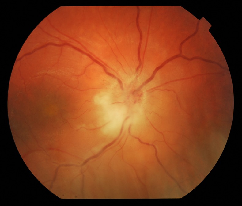
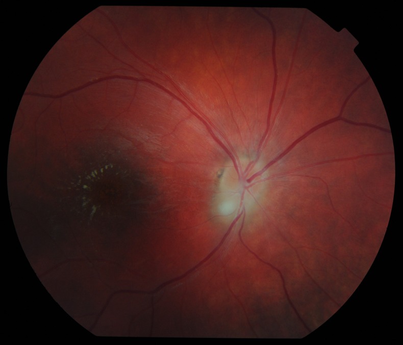
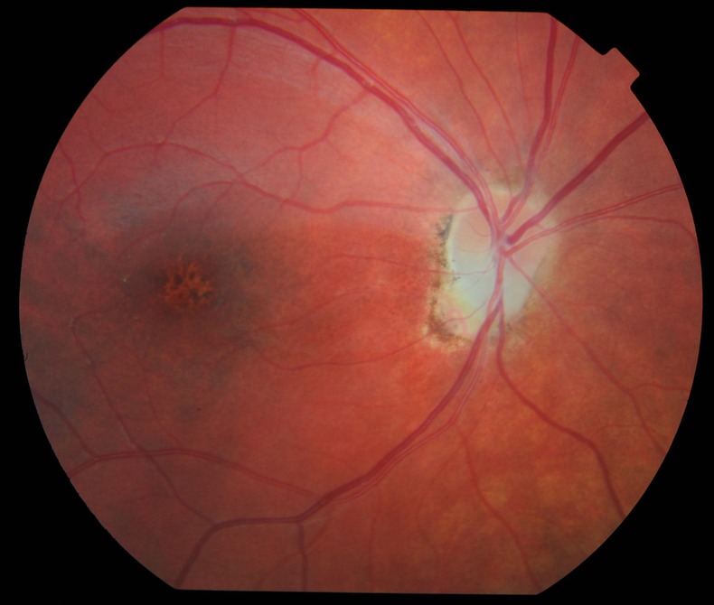
Toxoplasma is a leading cause of posterior uveitis in immunocompetent patients manifesting as a focal posterior retinochoroiditis. The clinical diagnosis of ocular toxoplasmosis is usually straightforward. There is typically a fluffy white retinal lesion which may lie adjacent to a pigmented chorioretinal scar and a prominent vitreous, or additionally, anterior chamber cellular reaction. Several unusual presentations in ocular toxoplasmosis have been reported, including: papillitis, neuroretinitis, retrobulbar neuritis, retinal detachment and macular oedema. This is a case of presumed primary toxoplasma papillitis in a 14-year-old child with complete absence of vitritis at presentation that made the diagnosis challenging. This evolved into neuroretinitis that resolved upon introducing antitoxoplasma antibiotics.
Figures
References
-
- Folk JC, Lobes LA. Presumed toxoplasmic papillitis. Ophthalmology 1984;2013:64–7 - PubMed
-
- Fish RH, Hoskins JC, Kline LB. Toxoplasma neuroretinitis. Ophthalmology 1993;2013:1177–82 - PubMed
-
- Rahi A, Tabarra K. Laboratory investigations in posterior uveitis. Int Ophthalmol Clin 1995;2013:65–6 - PubMed
-
- De Groot-Mijnes JD, Rothova A, Van Loon AM, et al. Polymerase chain reaction and Goldmann-Witmer coefficient analysis are complimentary for the diagnosis of infectious uveitis. Am J Ophthalmol 2006;2013:313–18 - PubMed
Publication types
MeSH terms
Substances
LinkOut - more resources
Full Text Sources
Other Literature Sources